Copyright
©The Author(s) 2018.
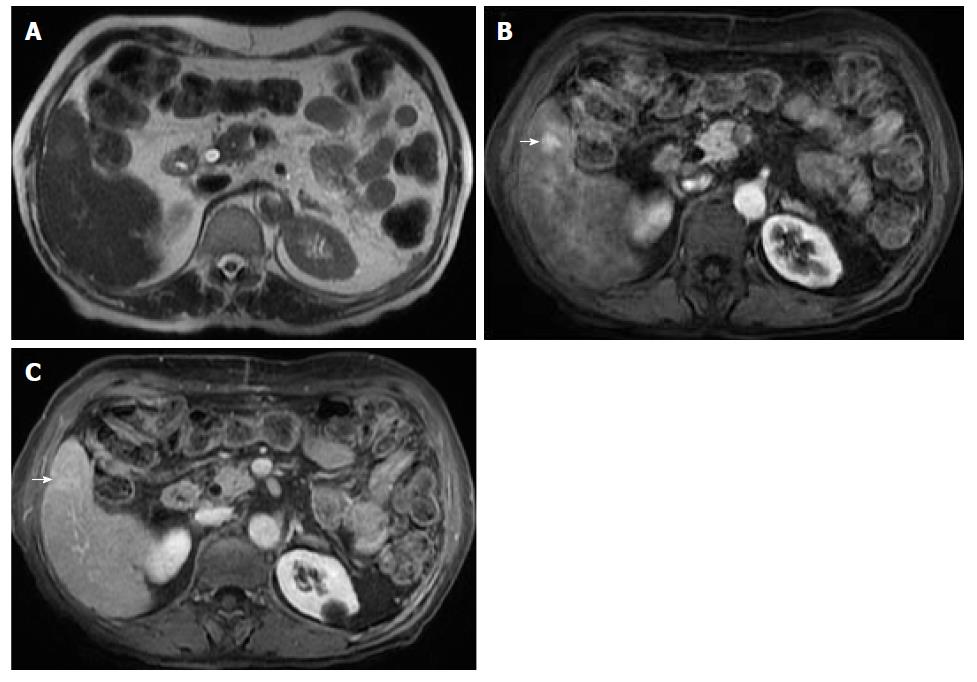
Figure 3 Small hepatocellular carcinoma without washout.
T2-weighted image (A), and post-contrast fat-suppressed 3D-GRE T1-weighted images during the late hepatic arterial (B) and delayed phase (C). A small nodule with mild signal increase on T2 weighed image is seen in the right liver lobe (A). It shows intense increased arterial hyperenhancement (arrow, B) and fading in subsequent phases without a clear washout in the delayed phase (C).
- Citation: Campos-Correia D, Cruz J, Matos AP, Figueiredo F, Ramalho M. Magnetic resonance imaging ancillary features used in Liver Imaging Reporting and Data System: An illustrative review. World J Radiol 2018; 10(2): 9-23
- URL: https://www.wjgnet.com/1949-8470/full/v10/i2/9.htm
- DOI: https://dx.doi.org/10.4329/wjr.v10.i2.9


