Copyright
©The Author(s) 2018.
World J Radiol. Nov 28, 2018; 10(11): 150-161
Published online Nov 28, 2018. doi: 10.4329/wjr.v10.i11.150
Published online Nov 28, 2018. doi: 10.4329/wjr.v10.i11.150
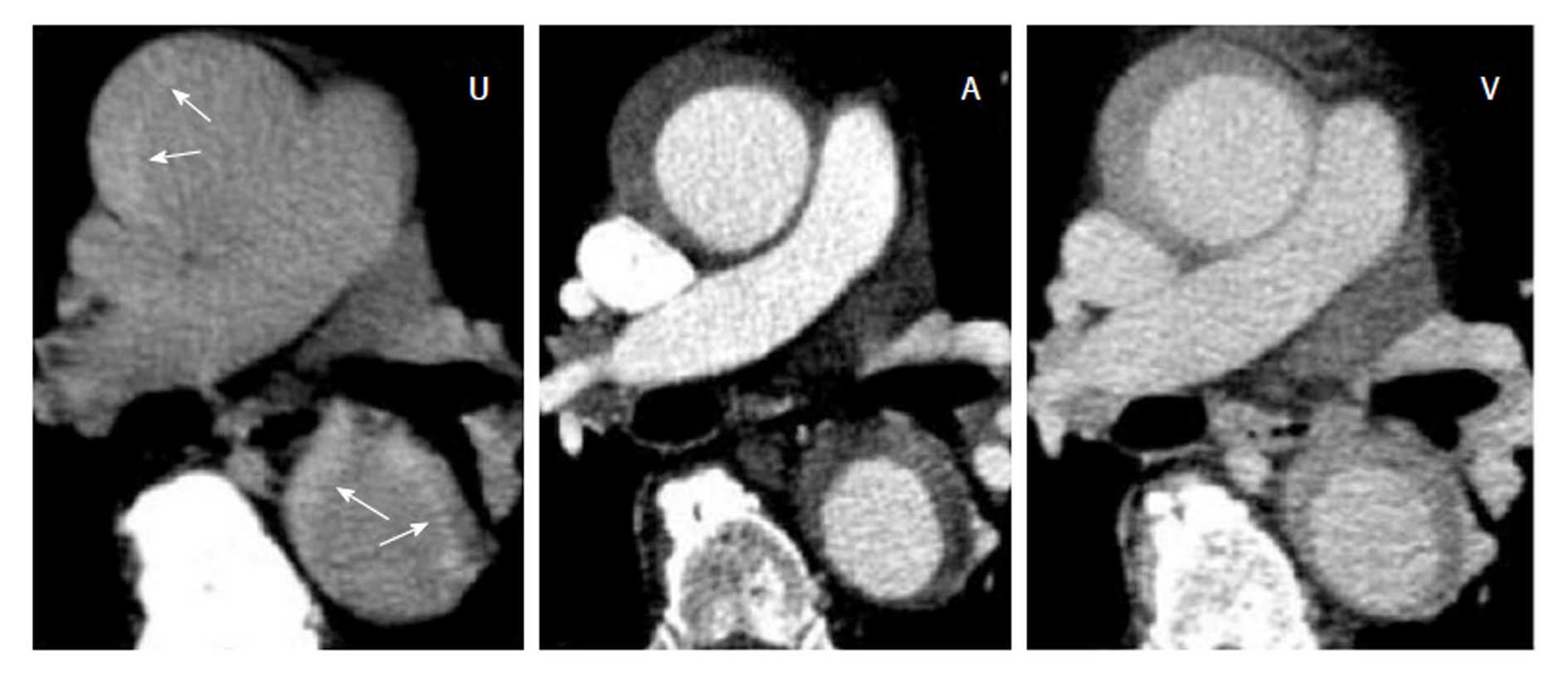
Figure 6 Acute intramural hematoma.
Triphasic computed tomography angiography with an acute intramural hematoma (IMH) type Stanford A in the ascending and descending aorta. The unenhanced scan (U) shows a hyperdense wall thickening compared to the lumen (arrows). In the arterial (A) and venous (V) phase of the enhanced scans, the IMH con not be differentiated from a thrombotic layer.
- Citation: Panagiotopoulos N, Drüschler F, Simon M, Vogt FM, Wolfrum S, Desch S, Richardt D, Barkhausen J, Hunold P. Significance of an additional unenhanced scan in computed tomography angiography of patients with suspected acute aortic syndrome. World J Radiol 2018; 10(11): 150-161
- URL: https://www.wjgnet.com/1949-8470/full/v10/i11/150.htm
- DOI: https://dx.doi.org/10.4329/wjr.v10.i11.150


