Copyright
©2009 Baishideng Publishing Group Co.
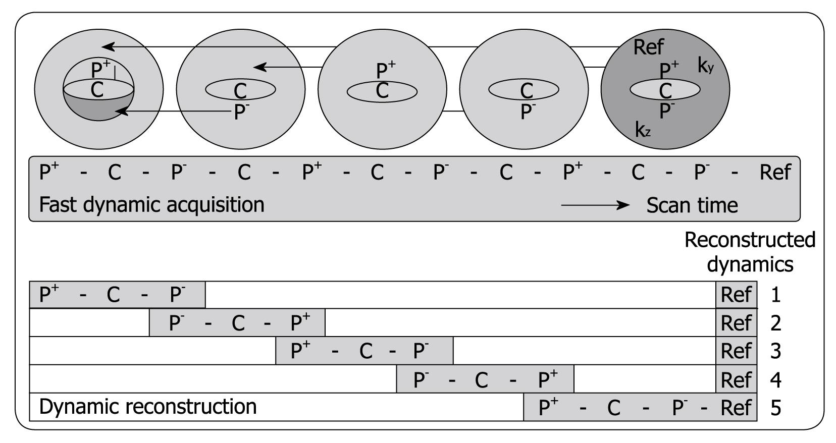
Figure 1 Schematic depiction of the alternating viewsharing technique.
The central ky-kz disk defined by the keyhole percentage is subdivided in 3 regions, P+, C and P-, where P+ and P- cover positive and negative peripheral regions in this central disk and C is the central region as shown. The central region C is acquired in each dynamic scan while regions P+ and P- are shared with subsequent dynamic scans according to an alternating viewsharing scheme: P+-C-P--C-P+-C-P--C-P+-Ref. The P- and P+ parts from subsequent keyhole scans are shared in the reconstruction process.
- Citation: Coenegrachts K. Magnetic resonance imaging of the liver: New imaging strategies for evaluating focal liver lesions. World J Radiol 2009; 1(1): 72-85
- URL: https://www.wjgnet.com/1949-8470/full/v1/i1/72.htm
- DOI: https://dx.doi.org/10.4329/wjr.v1.i1.72


