Copyright
©2009 Baishideng Publishing Group Co.
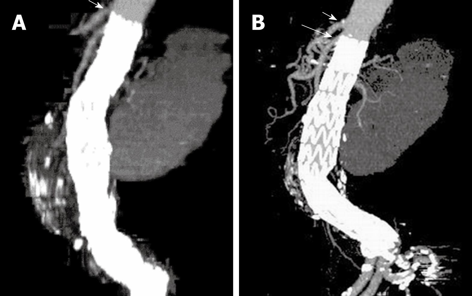
Figure 9 A stent graft migration of 10.
2 mm was noticed in a recent sagittal MIP image (B) in a patient treated with suprarenal stent graft 3 years ago (A). Short arrows indicate celiac axis, while long arrow refer to SMA.
- Citation: Sun Z. Endovascular stent graft repair of abdominal aortic aneurysms: Current status and future directions. World J Radiol 2009; 1(1): 63-71
- URL: https://www.wjgnet.com/1949-8470/full/v1/i1/63.htm
- DOI: https://dx.doi.org/10.4329/wjr.v1.i1.63


