Copyright
©2009 Baishideng Publishing Group Co.
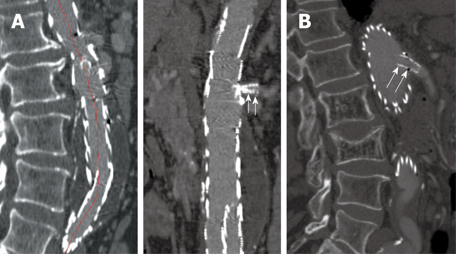
Figure 7 Curved planar reformatted images are produced following the centreline of the abdominal aorta to demonstrate the fenestrated renal stent (arrows in A) and the intra-aortic portion of fenestrated stent deployed into the SMA (arrows in B).
- Citation: Sun Z. Endovascular stent graft repair of abdominal aortic aneurysms: Current status and future directions. World J Radiol 2009; 1(1): 63-71
- URL: https://www.wjgnet.com/1949-8470/full/v1/i1/63.htm
- DOI: https://dx.doi.org/10.4329/wjr.v1.i1.63


