Copyright
©2009 Baishideng Publishing Group Co.
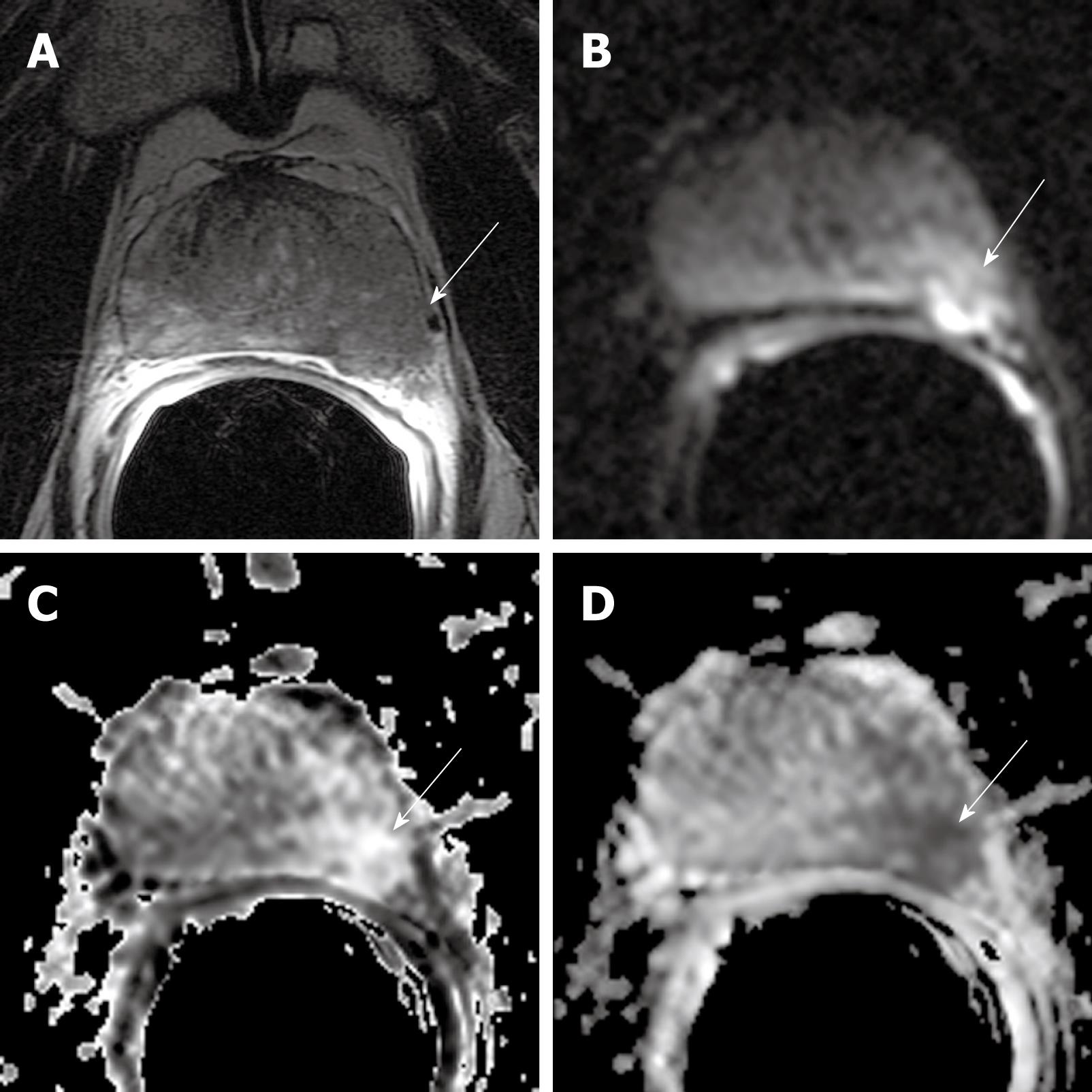
Figure 6 MR images of established ECE of prostate cancer into the periprostatic fat in a 59-year-old man with PSA level of 21.
7 ng/mL and Gleason grade 4 + 4 and PT3a. Transverse 3 mm-thick MR (5800/108) image (A) and transverse diffusion image (4000/85. b1000) (B), exponential ADC (C) and ADC (D) of the prostate mid-gland reveal an infiltrative peripheral zone tumor (arrows) that extends into the left periprostatic fat.
- Citation: Wang L. Incremental value of magnetic resonance imaging in the advanced management of prostate cancer. World J Radiol 2009; 1(1): 3-14
- URL: https://www.wjgnet.com/1949-8470/full/v1/i1/3.htm
- DOI: https://dx.doi.org/10.4329/wjr.v1.i1.3


