Copyright
©2009 Baishideng Publishing Group Co.
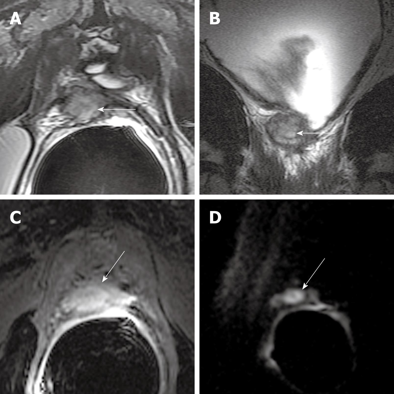
Figure 3 1.
5T MR images of locally recurrent prostate cancer in a 63-year-old man with rising PSA levels after radical prostatectomy. Transverse 3 mm-thick T2-WI (4000/125) (A) and coronal 3 mm-thick T2-WI (5300/100) (B) show intermediate SI mass (arrows) to the right posterior aspect of the bladder neck at the anastomosis; C: Transverse 3 mm-thick T1-WI (5.5/2.4) shows significant enhancement of the mass (arrow) after intravenous administration of gadolinium; D: Transverse 3 mm-thick diffusion-weighted image (DWI) (3500/93, b-value of 1000 s/mm2) shows intense increased signal (restricted diffusion) throughout the mass (arrow).
- Citation: Wang L. Incremental value of magnetic resonance imaging in the advanced management of prostate cancer. World J Radiol 2009; 1(1): 3-14
- URL: https://www.wjgnet.com/1949-8470/full/v1/i1/3.htm
- DOI: https://dx.doi.org/10.4329/wjr.v1.i1.3


