Copyright
©The Author(s) 2024.
World J Radiol. Apr 28, 2024; 16(4): 82-93
Published online Apr 28, 2024. doi: 10.4329/wjr.v16.i4.82
Published online Apr 28, 2024. doi: 10.4329/wjr.v16.i4.82
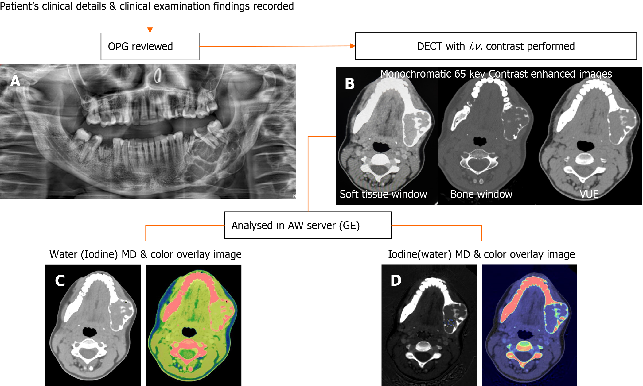
Figure 1 Workflow of patients undergoing dual-energy computed tomography Imaging.
A: Orthopantomogram images are reviewed first; B: Followed by dual-energy computed tomography (DECT) acquisition using intravenous non-ionic iodinated contrast; C: Water (Iodine) with color overlay; D: Iodine (water) with color overlay are the material density images reconstructed in the dedicated software for quantitative analysis. DECT: Dual-energy computed tomography; OPG: Orthopantomography; MD: Material decomposition.
Figure 2 Classification of lesions based on computed tomography morphology (density and locularity).
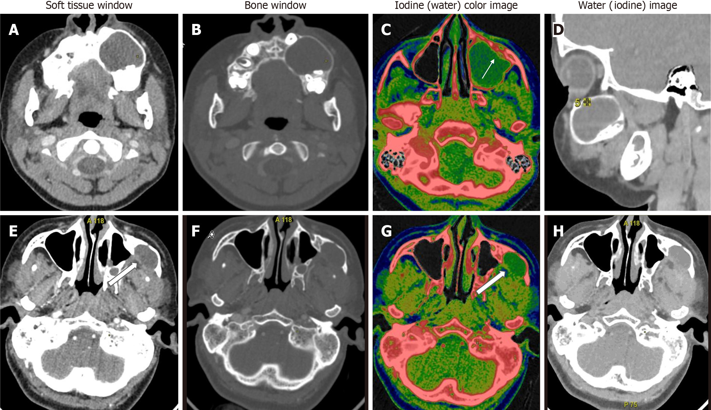
Figure 3 Unilocular lytic lesions differentiated based on water concentration.
A-D: Unilocular ameloblastoma - contrast-enhanced dual-energy computed tomography images show a well-defined lytic unilocular cystic, expansile lesion in the left maxilla with mild peripheral rim enhancement better appreciated on iodine colour overlay images (white arrow). Water (iodine) material decomposition (MD) images showed a WC of 986 μg/cm3 in the cystic component; E-H: Odontogenic keratocyst is also a well-defined lytic, unilocular cystic, expansile lesion in the left lateral wall of the maxillary sinus with a small enhancing mural component posteriorly (white open arrows). Water (Iodine) MD images revealed a water concentration of 1045 μg/cm3 in the cystic component. In this case, due to the paucity of soft tissue components, iodine concentration did not help; however, the water concentration of the cystic component differed significantly, aiding in the diagnosis.
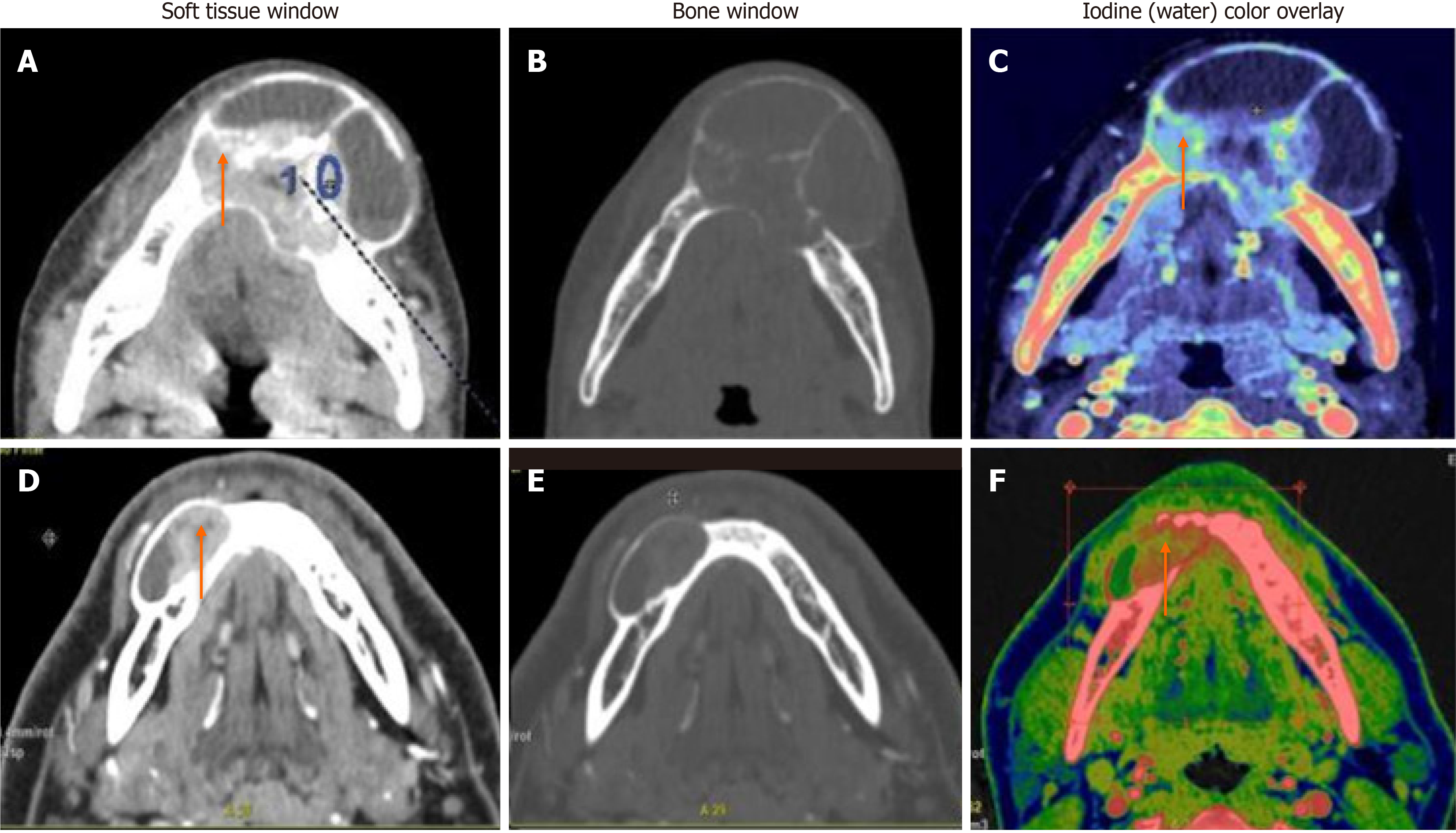
Figure 4 Multilocular solid-cystic lesions differentiated based on iodine concentration.
A-C: Central giant cell granuloma - A well-defined expansile multiloculated solid-cystic tumor in the left anterior mandible crossing the midline. Iodine images with a color overlay (C) showed an iodine concentration (IC) of 57 × 100 μg/cm3 of the solid enhancing part (orange arrow); D-F: Ameloblastoma - a well-defined lytic, unilocular, solid-cystic, expansile lesion with an enhancing soft tissue component (orange arrow). Iodine images with a color overlay (F) show increased iodine content (areas in red) within the soft tissue (IC of 23 × 100 μg/cm3).
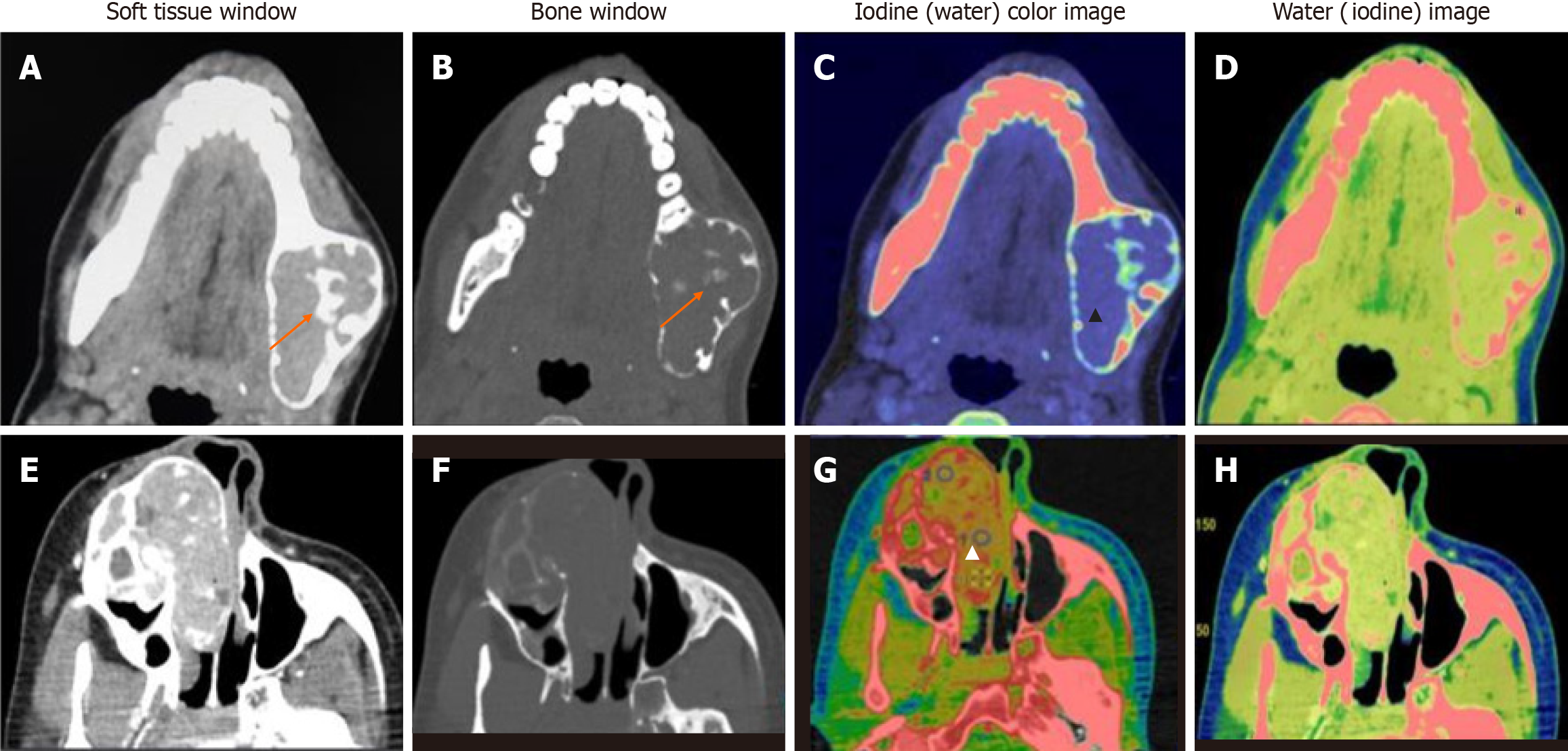
Figure 5 Mixed lytic-sclerotic lesions.
A-D: Central giant cell granuloma well-defined, mixed lytic-sclerotic buccolingual expansile lesion with a narrow zone of transition. Central ossific foci are seen (orange arrows). Iodine (water) material decomposition overlay images show a mild, homogeneous iodine concentration (blue region within the tumor - black arrowhead) (C). Water (Iodine) images show no cystic or necrotic areas (D); E-H: Ossifying fibroma well- defined expansile mass epicentered in the right maxilla, showing heterogeneous enhancement in its lytic soft tissue component with multiple sclerotic foci extending into the nasal cavity (E). The iodine image shows foci of increased iodine concentration (red areas - white arrowhead) (G). Lower iodine concentration and higher water concentration were seen in the latter, which suggested “other jaw tumor” as in this case.
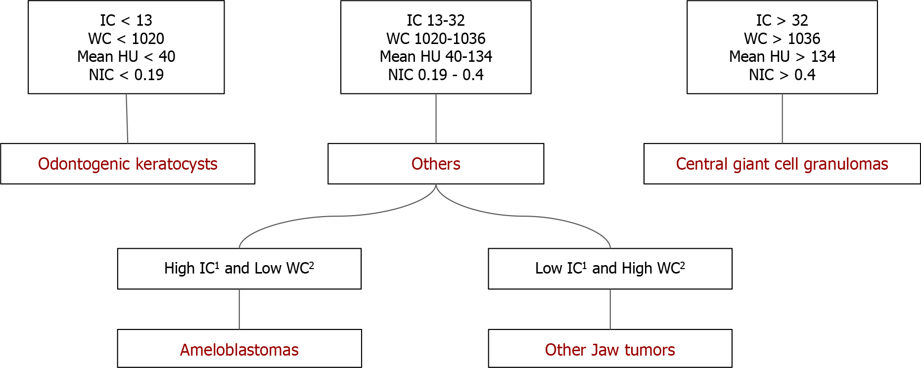
Figure 6 Flowchart showing algorithmic differentiation of jaw tumors in the study population based on the quantitative analysis.
Central giant cell granulomas had higher iodine concentration (IC), water concentration (WC), and normalized IC (NIC), odontogenic keratocysts with lower IC, WC, and NIC, ameloblastomas and other jaw tumor group showed values in between. Between the two, ameloblastomas had higher IC with lower WC while other jaw tumor group had lower IC with higher WC. 1Not significant; 2Marginally significant. WC: Water concentration; IC: Iodine concentration; NIC: Normalized iodine concentration.
- Citation: Viswanathan DJ, Bhalla AS, Manchanda S, Roychoudhury A, Mishra D, Mridha AR. Characterization of tumors of jaw: Additive value of contrast enhancement and dual-energy computed tomography. World J Radiol 2024; 16(4): 82-93
- URL: https://www.wjgnet.com/1949-8470/full/v16/i4/82.htm
- DOI: https://dx.doi.org/10.4329/wjr.v16.i4.82


