Copyright
©The Author(s) 2017.
World J Cardiol. Jul 26, 2017; 9(7): 600-608
Published online Jul 26, 2017. doi: 10.4330/wjc.v9.i7.600
Published online Jul 26, 2017. doi: 10.4330/wjc.v9.i7.600
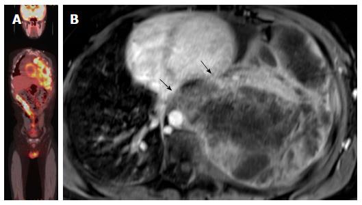
Figure 8 Evaluation of the aggressiveness of the lesion and assessment of cardiac involvement.
A: Evaluation of the aggressiveness of the lesion and assessment of cardiac involvement; whole body PET-CT image of patient with extensive Ewing sarcoma of the left hemithorax, PET-CT images are not sufficient to evaluate local extension of the tumor to the heart; B: Axial delayed enhancement image shows large necrotic mass occupying the left hemithorax with direct left ventricle (upper arrow) the arrow without circle and left atrial invasion (lower arrow). PET: Positron emission tomography; CT: Computed tomography.
- Citation: Fathala A, Abouzied M, AlSugair AA. Cardiac and pericardial tumors: A potential application of positron emission tomography-magnetic resonance imaging. World J Cardiol 2017; 9(7): 600-608
- URL: https://www.wjgnet.com/1949-8462/full/v9/i7/600.htm
- DOI: https://dx.doi.org/10.4330/wjc.v9.i7.600


