Copyright
©The Author(s) 2017.
World J Cardiol. Jul 26, 2017; 9(7): 600-608
Published online Jul 26, 2017. doi: 10.4330/wjc.v9.i7.600
Published online Jul 26, 2017. doi: 10.4330/wjc.v9.i7.600
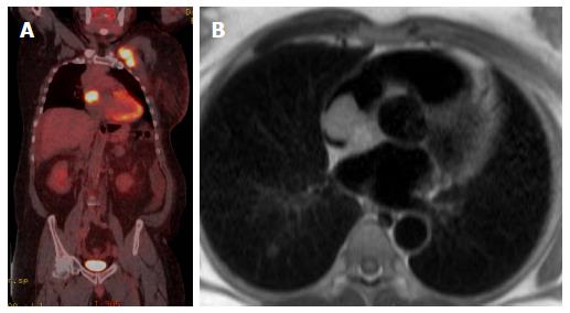
Figure 7 M staging.
A: M staging: Coronal PET-CT image of patient with lymphoma with unexpected cardiac involvement; B: Axial T1-weighted image at the level of interatrial septum shows well defined mass attached to the atrial septum and nearly fills the right atrium and was proved to be lymphoma. PET: Positron emission tomography; CT: Computed tomography.
- Citation: Fathala A, Abouzied M, AlSugair AA. Cardiac and pericardial tumors: A potential application of positron emission tomography-magnetic resonance imaging. World J Cardiol 2017; 9(7): 600-608
- URL: https://www.wjgnet.com/1949-8462/full/v9/i7/600.htm
- DOI: https://dx.doi.org/10.4330/wjc.v9.i7.600


