Copyright
©The Author(s) 2017.
World J Cardiol. Jul 26, 2017; 9(7): 600-608
Published online Jul 26, 2017. doi: 10.4330/wjc.v9.i7.600
Published online Jul 26, 2017. doi: 10.4330/wjc.v9.i7.600
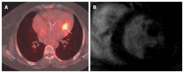
Figure 1 Localization of abnormal fluorodeoxygluocse activity in the heart.
A: Axial fused PET-CT image shows focal intense FDG uptake in the heart (arrow) in patient with melanoma that thought represents cardiac metastases; B: Selected delayed enhancement short axis image of the same patient with other images (not shown) shows no abnormal enhancement or tissue infiltration, the FDG uptake was corresponding to hypertrophic papillary muscle, follow PET-CT was normal. PET: Positron emission tomography; CT: Computed tomography; FDG: Fluorodeoxygluocse.
- Citation: Fathala A, Abouzied M, AlSugair AA. Cardiac and pericardial tumors: A potential application of positron emission tomography-magnetic resonance imaging. World J Cardiol 2017; 9(7): 600-608
- URL: https://www.wjgnet.com/1949-8462/full/v9/i7/600.htm
- DOI: https://dx.doi.org/10.4330/wjc.v9.i7.600


