Copyright
©The Author(s) 2017.
World J Cardiol. Apr 26, 2017; 9(4): 384-390
Published online Apr 26, 2017. doi: 10.4330/wjc.v9.i4.384
Published online Apr 26, 2017. doi: 10.4330/wjc.v9.i4.384
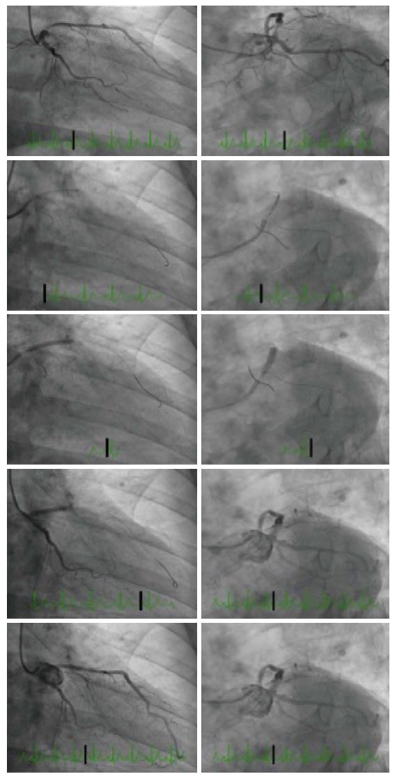
Figure 3 Angiographic images for case 2.
Coronary angiography showed separate ostia of the LAD and left circumflex artery (LCx) and proximal moderate to severe LAD stenosis. RAO caudal (left) and LAO caudal (right) images with diagnostic images (top), Szabo technique (center three panels) and final images (bottom). LAD: Left anterior descending; LCx: Left circumflex artery.
- Citation: Yu K, Hundal H, Zynda T, Seto A. Three-dimensional optical coherence tomography reconstruction of bifurcation stenting using the Szabo anchor-wire technique. World J Cardiol 2017; 9(4): 384-390
- URL: https://www.wjgnet.com/1949-8462/full/v9/i4/384.htm
- DOI: https://dx.doi.org/10.4330/wjc.v9.i4.384


