Copyright
©The Author(s) 2017.
World J Cardiol. Mar 26, 2017; 9(3): 268-276
Published online Mar 26, 2017. doi: 10.4330/wjc.v9.i3.268
Published online Mar 26, 2017. doi: 10.4330/wjc.v9.i3.268
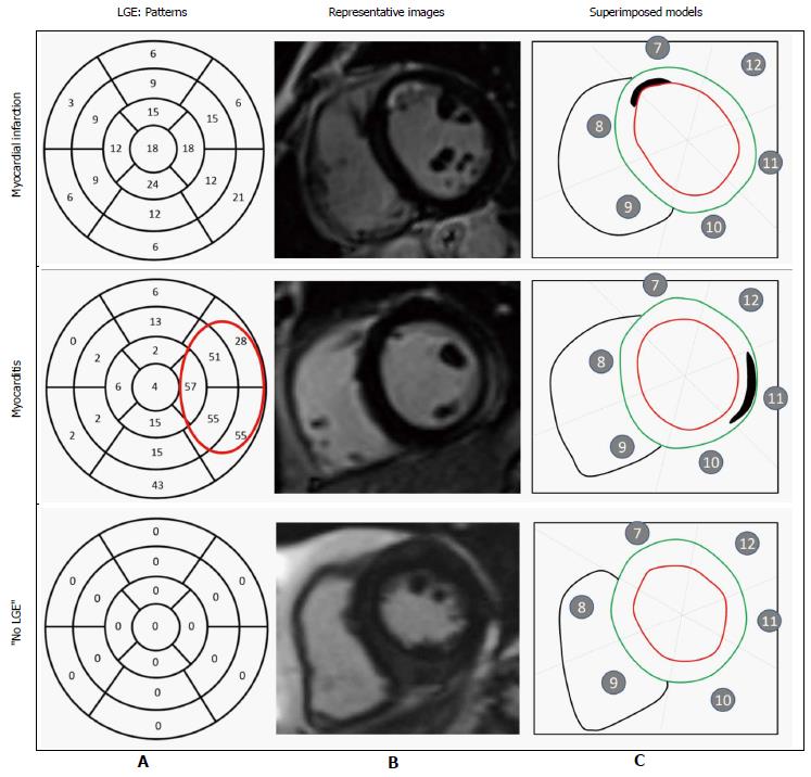
Figure 2 Pattern of late gadolinium enhancement.
A: Incidence of late gadolinium enhancement (LGE) within each segment (percentage) of all patients, including those with and without LGE. In myocarditis cases, LGE was predominantly on the lateral wall. In myocardial infarction, no specific pattern of LGE could be identified; B: Representative short-axis slice images showing LGE location; C: Superimposed segmental models showing location and spatial extent of LGE (outlined).
- Citation: Bière L, Niro M, Pouliquen H, Gourraud JB, Prunier F, Furber A, Probst V. Risk of ventricular arrhythmia in patients with myocardial infarction and non-obstructive coronary arteries and normal ejection fraction. World J Cardiol 2017; 9(3): 268-276
- URL: https://www.wjgnet.com/1949-8462/full/v9/i3/268.htm
- DOI: https://dx.doi.org/10.4330/wjc.v9.i3.268


