Copyright
©The Author(s) 2017.
World J Cardiol. Feb 26, 2017; 9(2): 92-108
Published online Feb 26, 2017. doi: 10.4330/wjc.v9.i2.92
Published online Feb 26, 2017. doi: 10.4330/wjc.v9.i2.92
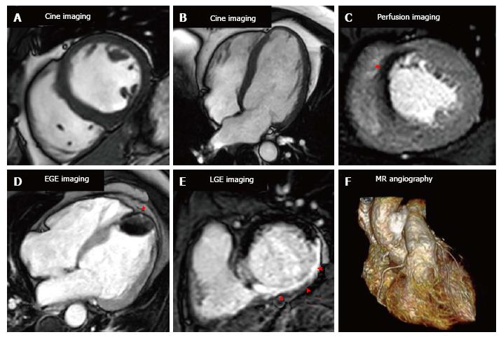
Figure 1 Cardiovascular magnetic resonance imaging techniques.
A and B show short axis and 4 chamber cine images respectively for anatomical and functional assessment; C shows stress perfusion with a septal perfusion defect (arrow); D shows early gadolinium enhancement imaging with a large apical thrombus (arrow); E is late gadolinium enhanced imaging with a transmural inferior infarction (arrows); F is 3D whole heart magnetic resonance angiography. LGE: Late gadolinium enhancement; EGE: Early gadolinium enhancement.
- Citation: Foley JRJ, Plein S, Greenwood JP. Assessment of stable coronary artery disease by cardiovascular magnetic resonance imaging: Current and emerging techniques. World J Cardiol 2017; 9(2): 92-108
- URL: https://www.wjgnet.com/1949-8462/full/v9/i2/92.htm
- DOI: https://dx.doi.org/10.4330/wjc.v9.i2.92


