Copyright
©The Author(s) 2017.
World J Cardiol. Feb 26, 2017; 9(2): 191-195
Published online Feb 26, 2017. doi: 10.4330/wjc.v9.i2.191
Published online Feb 26, 2017. doi: 10.4330/wjc.v9.i2.191
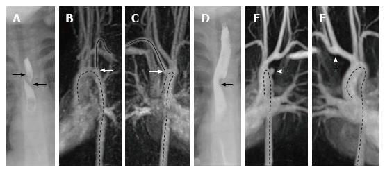
Figure 1 Fluoroscopy and magnetic resonance angiography at initial presentation and at follow-up.
A-C: Fluoroscopy and MRA at initial presentation; A: The arrows mark the outer boundary of the esophagus. There is a filling defect in between which runs from right side superior to left side inferior due to compression of the vessel; B and C: The arrow marks the right sided subclavian artery which originates distally to the left supraaortic vessels. The course of the artery is shown by the uninterrupted line. The dotted lines mark the course of the thoracic aorta; D-F: Fluoroscopy and MRA at follow up; D: The arrow marks a filling defect of the esophagus; E: The arrow marks the vascular stump. The dotted lines mark the course of the thoracic aorta; F: The arrow marks the anastomosis. The dotted line represents the course of the aorta. MRA: Magnetic resonance angiography.
- Citation: Mayer J, van der Werf-Grohmann N, Kroll J, Spiekerkoetter U, Stiller B, Grohmann J. Dysphagia after arteria lusoria dextra surgery: Anatomical considerations before redo-surgery. World J Cardiol 2017; 9(2): 191-195
- URL: https://www.wjgnet.com/1949-8462/full/v9/i2/191.htm
- DOI: https://dx.doi.org/10.4330/wjc.v9.i2.191


