Copyright
©The Author(s) 2017.
World J Cardiol. Feb 26, 2017; 9(2): 109-133
Published online Feb 26, 2017. doi: 10.4330/wjc.v9.i2.109
Published online Feb 26, 2017. doi: 10.4330/wjc.v9.i2.109
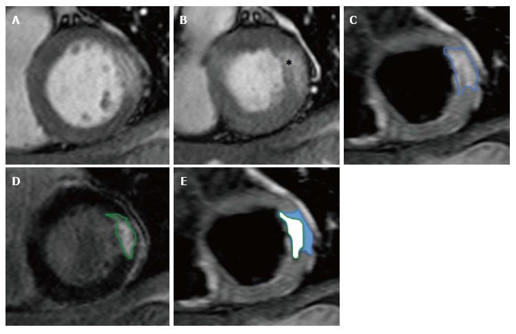
Figure 8 Calculation of salvaged myocardium.
A: SSFP end-diastolic cine image; B: SSFP end-systolic cine image showing hypokinetic basal anterolateral segment (*); C: T2w-STIR image showing oedema (AAR) in anterolateral wall consistent with circumflex artery occlusion; D: Corresponding LGE image with near-transmural infarction; E: Calculation of salvaged myocardium in blue. SSFP: Steady-state free precession; T2w-STIR: T2-weighted short-tau inversion-recovery sequence; LGE: Late gadolinium enhancement.
- Citation: Khan JN, McCann GP. Cardiovascular magnetic resonance imaging assessment of outcomes in acute myocardial infarction. World J Cardiol 2017; 9(2): 109-133
- URL: https://www.wjgnet.com/1949-8462/full/v9/i2/109.htm
- DOI: https://dx.doi.org/10.4330/wjc.v9.i2.109


