Copyright
©The Author(s) 2017.
World J Cardiol. Feb 26, 2017; 9(2): 109-133
Published online Feb 26, 2017. doi: 10.4330/wjc.v9.i2.109
Published online Feb 26, 2017. doi: 10.4330/wjc.v9.i2.109
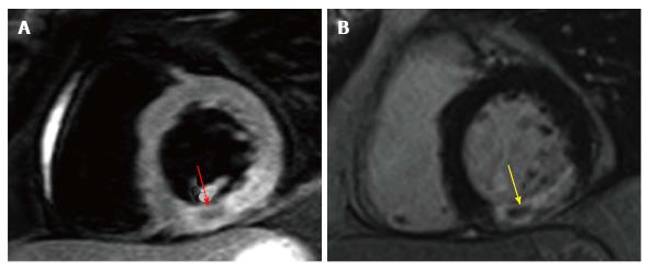
Figure 6 Intramyocardial haemorrhage on cardiovascular magnetic resonance.
A: T2-weighted spin-echo image with hypointensity corresponding with IMH within the hyperintense oedematous region in the inferior wall (red arrow); B: Corresponding LGE image showing co-localisation of IMH and L-MVO (yellow arrow). IMH: Intramyocardial haemorrhage; LGE: Late gadolinium enhancement; MVO: Microvascular obstruction.
- Citation: Khan JN, McCann GP. Cardiovascular magnetic resonance imaging assessment of outcomes in acute myocardial infarction. World J Cardiol 2017; 9(2): 109-133
- URL: https://www.wjgnet.com/1949-8462/full/v9/i2/109.htm
- DOI: https://dx.doi.org/10.4330/wjc.v9.i2.109


