Copyright
©The Author(s) 2017.
World J Cardiol. Feb 26, 2017; 9(2): 109-133
Published online Feb 26, 2017. doi: 10.4330/wjc.v9.i2.109
Published online Feb 26, 2017. doi: 10.4330/wjc.v9.i2.109
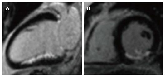
Figure 4 Late gadolinium enhancement of acute infarct.
Infarct appears white (enhanced) in the inferior wall, with unaffected myocardium black (nulled). A: 2-chamber long-axis view; B: Short-axis view, mid ventricular level. The posteromedial papillary muscle is also infarcted in the short-axis view.
- Citation: Khan JN, McCann GP. Cardiovascular magnetic resonance imaging assessment of outcomes in acute myocardial infarction. World J Cardiol 2017; 9(2): 109-133
- URL: https://www.wjgnet.com/1949-8462/full/v9/i2/109.htm
- DOI: https://dx.doi.org/10.4330/wjc.v9.i2.109


