Copyright
©The Author(s) 2017.
World J Cardiol. Feb 26, 2017; 9(2): 109-133
Published online Feb 26, 2017. doi: 10.4330/wjc.v9.i2.109
Published online Feb 26, 2017. doi: 10.4330/wjc.v9.i2.109
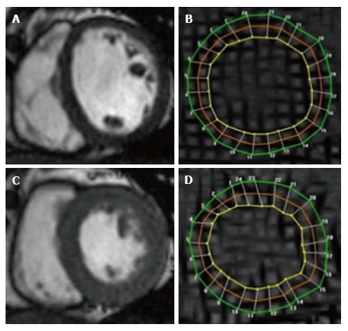
Figure 2 Cardiovascular magnetic resonance assessment of strain using tissue tagging.
Cine SSFP images in end-diastole (A) and end-systole (C), with corresponding Spatial Modulation of Motion (SPAMM) tagged images (B and D). Grid lines (tags) are visible and contours drawn at 3 myocardial levels [green (epicardial), red (mid myocardial), yellow (endocardial)] allow tracking of myocardial motion and strain (circumferential), here using Harmonic Phase Analysis.
- Citation: Khan JN, McCann GP. Cardiovascular magnetic resonance imaging assessment of outcomes in acute myocardial infarction. World J Cardiol 2017; 9(2): 109-133
- URL: https://www.wjgnet.com/1949-8462/full/v9/i2/109.htm
- DOI: https://dx.doi.org/10.4330/wjc.v9.i2.109


