Copyright
©The Author(s) 2017.
World J Cardiol. Dec 26, 2017; 9(12): 842-847
Published online Dec 26, 2017. doi: 10.4330/wjc.v9.i12.842
Published online Dec 26, 2017. doi: 10.4330/wjc.v9.i12.842
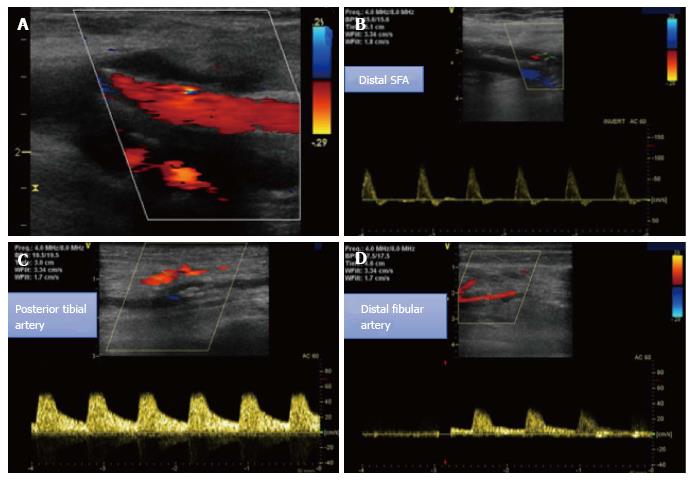
Figure 4 Duplex sonography at follow-up.
A, B: Well perfused SFA with biphasic flow in the distal SFA and in the popliteal artery; C, D: Monophasic flow in the distal posterior tibial and fibular arteries. SFA: Superficial femoral artery.
- Citation: Korosoglou G, Eisele T, Raupp D, Eisenbach C, Giusca S. Successful recanalization of long femoro-crural occlusive disease after failed bypass surgery. World J Cardiol 2017; 9(12): 842-847
- URL: https://www.wjgnet.com/1949-8462/full/v9/i12/842.htm
- DOI: https://dx.doi.org/10.4330/wjc.v9.i12.842


