Copyright
©The Author(s) 2017.
World J Cardiol. Dec 26, 2017; 9(12): 842-847
Published online Dec 26, 2017. doi: 10.4330/wjc.v9.i12.842
Published online Dec 26, 2017. doi: 10.4330/wjc.v9.i12.842
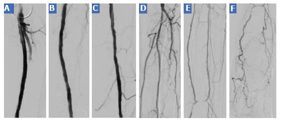
Figure 3 Digital subtraction angiography in the second interventional session.
A, B: DSA images of the SFA; C: DSA image of popliteal artery; D-F: DSA images of crural and foot arteries after the second angiographic procedure. SFA: Superficial femoral artery; DSA: Digital subtraction angiography.
- Citation: Korosoglou G, Eisele T, Raupp D, Eisenbach C, Giusca S. Successful recanalization of long femoro-crural occlusive disease after failed bypass surgery. World J Cardiol 2017; 9(12): 842-847
- URL: https://www.wjgnet.com/1949-8462/full/v9/i12/842.htm
- DOI: https://dx.doi.org/10.4330/wjc.v9.i12.842


