Copyright
©The Author(s) 2017.
World J Cardiol. Oct 26, 2017; 9(10): 773-786
Published online Oct 26, 2017. doi: 10.4330/wjc.v9.i10.773
Published online Oct 26, 2017. doi: 10.4330/wjc.v9.i10.773
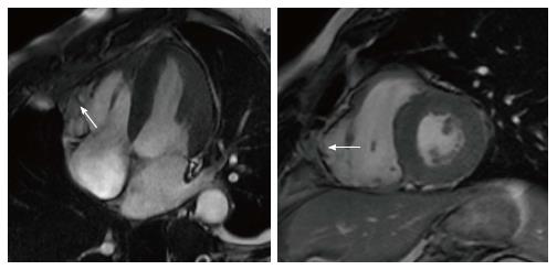
Figure 3 Subtricuspid involvement in arrhythmogenic right ventricular cardiomyopathy.
Dilated right ventricle with bulging of the subtricuspid region (arrow). The right ventricular apex is relatively spared.
- Citation: De Maria E, Aldrovandi A, Borghi A, Modonesi L, Cappelli S. Cardiac magnetic resonance imaging: Which information is useful for the arrhythmologist? World J Cardiol 2017; 9(10): 773-786
- URL: https://www.wjgnet.com/1949-8462/full/v9/i10/773.htm
- DOI: https://dx.doi.org/10.4330/wjc.v9.i10.773


