Copyright
©The Author(s) 2017.
World J Cardiol. Oct 26, 2017; 9(10): 773-786
Published online Oct 26, 2017. doi: 10.4330/wjc.v9.i10.773
Published online Oct 26, 2017. doi: 10.4330/wjc.v9.i10.773
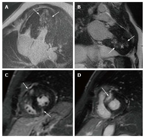
Figure 2 Late gadolinium enhancement patterns in hypertrophic cardiomyopathy patients.
A: LGE in the lateral wall (small arrows) and in the interventricular ventricular septum; B: LGE in the LV apex and inferior wall; C: LGE localized to the insertion area of the RV wall into the anterior and posterior ventricular septum; D: Transmural LGE involving the ventricular septum. LGE: Late gadolinium enhancement.
- Citation: De Maria E, Aldrovandi A, Borghi A, Modonesi L, Cappelli S. Cardiac magnetic resonance imaging: Which information is useful for the arrhythmologist? World J Cardiol 2017; 9(10): 773-786
- URL: https://www.wjgnet.com/1949-8462/full/v9/i10/773.htm
- DOI: https://dx.doi.org/10.4330/wjc.v9.i10.773


