Copyright
©The Author(s) 2016.
World J Cardiol. Sep 26, 2016; 8(9): 534-546
Published online Sep 26, 2016. doi: 10.4330/wjc.v8.i9.534
Published online Sep 26, 2016. doi: 10.4330/wjc.v8.i9.534
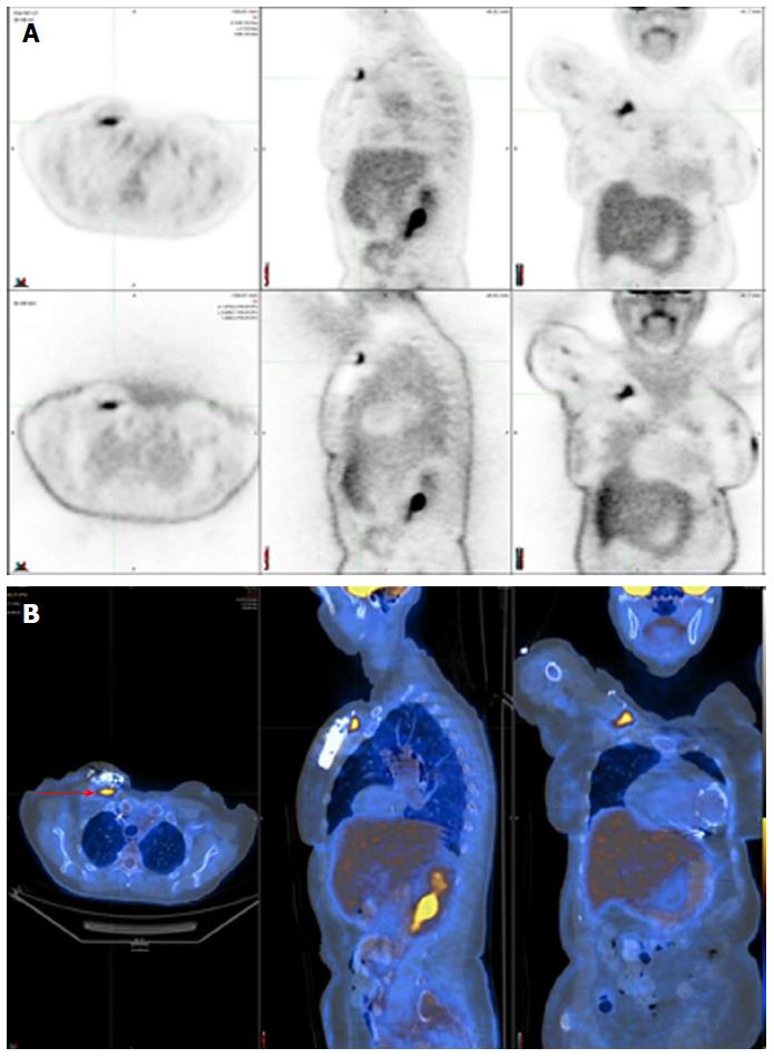
Figure 3 Positive 18F-fluorodeoxyglucose positron emission tomography/computed tomography in a patient with a deep pocket infection shown by focal 18F-fluorodeoxyglucose uptake just underneath the generator (red arrow).
A: SPECT displayed as transverse, sagittal, and coronal attenuation corrected (top row) and uncorrected images (bottom row); B: Hybrid SPECT/CT displayed as transverse, sagittal, and coronal images. SPECT/CT: Single-photon emission computed tomography/computed tomography.
- Citation: Sarrazin JF, Philippon F, Trottier M, Tessier M. Role of radionuclide imaging for diagnosis of device and prosthetic valve infections. World J Cardiol 2016; 8(9): 534-546
- URL: https://www.wjgnet.com/1949-8462/full/v8/i9/534.htm
- DOI: https://dx.doi.org/10.4330/wjc.v8.i9.534


