Copyright
©The Author(s) 2016.
World J Cardiol. Sep 26, 2016; 8(9): 520-533
Published online Sep 26, 2016. doi: 10.4330/wjc.v8.i9.520
Published online Sep 26, 2016. doi: 10.4330/wjc.v8.i9.520
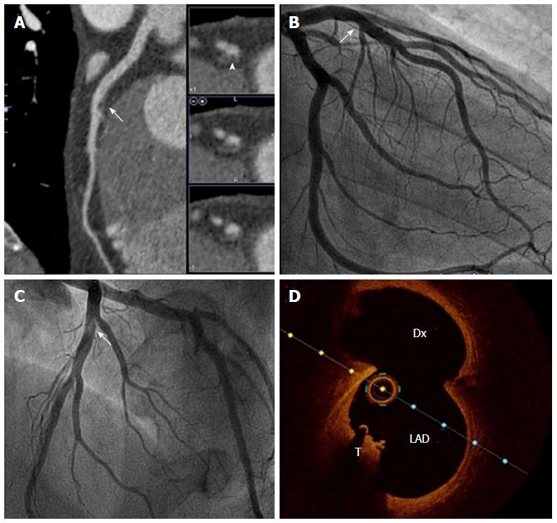
Figure 4 Coronary computed tomography characterization of plaque components.
Multimodal evaluation of a mid-LAD lesion in bifurcation with a Dx branch. A: CCT multiplanar reconstruction demonstrates a nonsignificant luminal narrowing in the mid LAD (arrow), and when short axis was evaluated the lesion fulfills noncalcified plaque features (arrowhead); B and C: ICA: The same nonobstructive lesion is observed in mid-LAD (arrow), which seems hyperlucent on LAO cranial projection (C); D: OCT confirms the presence of a red intracoronary thrombus (T) in the same location. CCT: Coronary computed tomography; LAD: Left anterior descending artery; Dx: Diagonal branch; ICA: Invasive coronary angiography; OCT: Optical coherence tomography.
- Citation: Pozo E, Agudo-Quilez P, Rojas-González A, Alvarado T, Olivera MJ, Jiménez-Borreguero LJ, Alfonso F. Noninvasive diagnosis of vulnerable coronary plaque. World J Cardiol 2016; 8(9): 520-533
- URL: https://www.wjgnet.com/1949-8462/full/v8/i9/520.htm
- DOI: https://dx.doi.org/10.4330/wjc.v8.i9.520


