Copyright
©The Author(s) 2016.
World J Cardiol. Sep 26, 2016; 8(9): 520-533
Published online Sep 26, 2016. doi: 10.4330/wjc.v8.i9.520
Published online Sep 26, 2016. doi: 10.4330/wjc.v8.i9.520
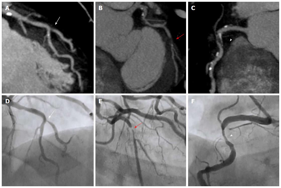
Figure 1 Coronary computed tomography stenosis evaluation compared with invasive coronary angiography.
Case of a patient with 3-vessel disease. Maximum intensity projection CCT findings are shown in the upper row with the corresponding ICA projections in the lower row. (A) demonstrates a significant stenosis in the ostium of the diagonal branch (arrow) at the level of its take-off from the mid-LAD in both CCT and ICA (D); In (B) CCT shows a subtotal occlusion in the proximal LCx (red arrow) that corresponds to a critical lesion at the same level in ICA (E); In CCT image from (C) a mixed plaque is detected in proximal RCA causing a significant stenosis (arrowhead), as corroborated by ICA (F). CCT: Coronary computed tomography; ICA: Invasive coronary angiography; LAD: Left anterior descending coronary artery; LCx: Left circumflex coronary artery; RCA: Right coronary artery.
- Citation: Pozo E, Agudo-Quilez P, Rojas-González A, Alvarado T, Olivera MJ, Jiménez-Borreguero LJ, Alfonso F. Noninvasive diagnosis of vulnerable coronary plaque. World J Cardiol 2016; 8(9): 520-533
- URL: https://www.wjgnet.com/1949-8462/full/v8/i9/520.htm
- DOI: https://dx.doi.org/10.4330/wjc.v8.i9.520


