Copyright
©The Author(s) 2016.
World J Cardiol. Sep 26, 2016; 8(9): 496-503
Published online Sep 26, 2016. doi: 10.4330/wjc.v8.i9.496
Published online Sep 26, 2016. doi: 10.4330/wjc.v8.i9.496
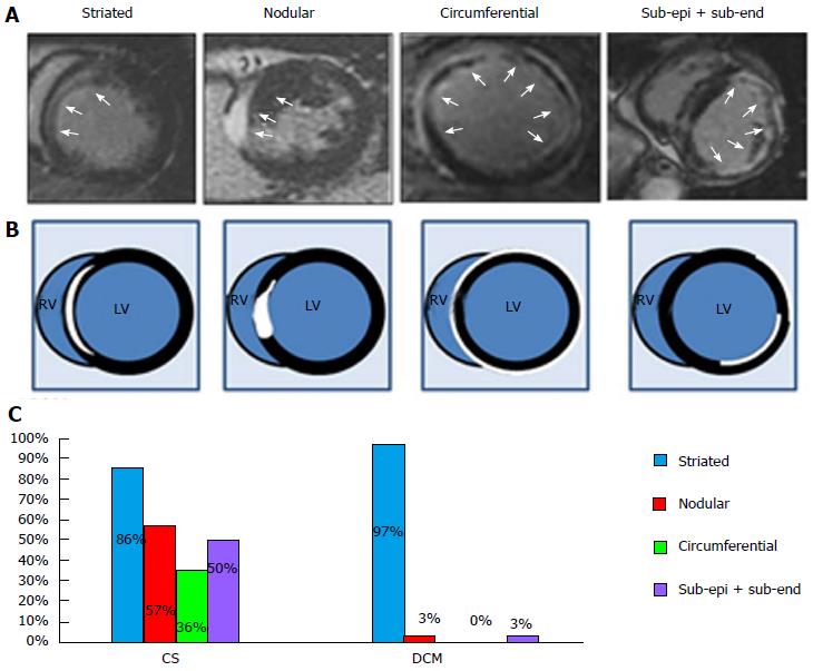
Figure 3 Typical late gadolinium enhancement distribution profiles.
Characteristic patterns of LE distribution in LE-MRI (A) and the cartoons (B). Striated: Striated LE distribution in midwall; Nodular: Nodular (transmural) LE distribution; Circumferential: Subepicardial LE distribution in > 50% circumferential LV wall; Sub-epi + sub-end: Subepicardial and subendocardial LE distribution with spared midwall (white arrows); C: The prevalence of characteristic patterns of LE distribution in patients with CS and with DCM. CS: Cardiac sarcoidosis; DCM: Dilated cardiomyopathy; LE: Late gadolinium enhancement; LV/RV: Left and right ventricles; MRI: Magnetic resonance imaging.
- Citation: Sano M, Satoh H, Suwa K, Saotome M, Urushida T, Katoh H, Hayashi H, Saitoh T. Intra-cardiac distribution of late gadolinium enhancement in cardiac sarcoidosis and dilated cardiomyopathy. World J Cardiol 2016; 8(9): 496-503
- URL: https://www.wjgnet.com/1949-8462/full/v8/i9/496.htm
- DOI: https://dx.doi.org/10.4330/wjc.v8.i9.496


