Copyright
©The Author(s) 2016.
World J Cardiol. Sep 26, 2016; 8(9): 496-503
Published online Sep 26, 2016. doi: 10.4330/wjc.v8.i9.496
Published online Sep 26, 2016. doi: 10.4330/wjc.v8.i9.496
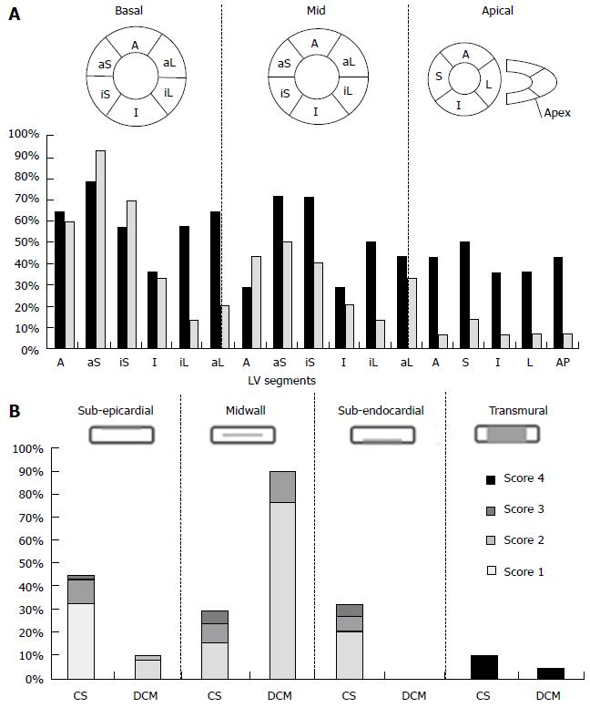
Figure 2 Intra-left ventricles (A) and intra-mural (B) late gadolinium enhancement distribution in patients with cardiac sarcoidosis and with dilated cardiomyopathy.
A: Columns indicate prevalence of LE at each LV segment in patients with CS (black) and with DCM (gray). A: Anterior; aL: Antero-lateral; aS: Anterior septal; I: Inferior; iL: Infero-lateral wall in basal, mid and apical LV; AP: LV apex; B: Columns consist of prevalence of LE with scores 1 to 3 at different intra-mural distribution in patients with CS and with DCM. Score 4 indicates the transmural distribution. CS: Cardiac sarcoidosis; DCM: Dilated cardiomyopathy; LV: Left ventricles.
- Citation: Sano M, Satoh H, Suwa K, Saotome M, Urushida T, Katoh H, Hayashi H, Saitoh T. Intra-cardiac distribution of late gadolinium enhancement in cardiac sarcoidosis and dilated cardiomyopathy. World J Cardiol 2016; 8(9): 496-503
- URL: https://www.wjgnet.com/1949-8462/full/v8/i9/496.htm
- DOI: https://dx.doi.org/10.4330/wjc.v8.i9.496


