Copyright
©The Author(s) 2016.
World J Cardiol. Jul 26, 2016; 8(7): 401-412
Published online Jul 26, 2016. doi: 10.4330/wjc.v8.i7.401
Published online Jul 26, 2016. doi: 10.4330/wjc.v8.i7.401
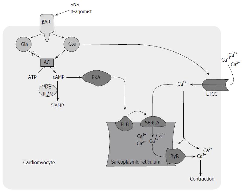
Figure 1 Beta-adrenoreceptor mediated signal transduction leads to the activation of both G stimulatory alpha protein and G inhibitory alpha protein.
Activated Gαs activates adenylyl cyclase (AC) which converts ATP into cAMP while activated Gαi inhibits AC. Activated Gαs also leads to calcium (Ca2+) mobilization into cardiomyocyte by activating L-type calcium channel (LTCC) independent of AC. This increase in intracellular Ca2+ concentration leads to activation of ryanodine receptor (RyR) which causes further release of Ca2+ from SR, a phenomenon known as calcium-induced calcium release. Elevated cAMP activates phosphokinase A (PKA) that inhibits phospholamban (PLB) by phosphorylating it. Phosphorylation of PLB increases uptake of Ca2+ from cytosol into the SR through sarcoplasmic reticulum calcium ATPase (SERCA). This enhanced Ca2+ entry into SR has positive impact on both systolic and diastolic function. In diastole, decreased intracellular Ca2+ causes relaxation. In systole increased release of Ca2+ from SR store through RyR activation increases inotropy. In the failing myocardium, chronic stimulation of βAR results in ineffective activation of AC, persistent activation of L-type calcium channel that increases Ca2+ influx, and decreased Ca2+ uptake into the SR due to decreased SERCA activity. This translates into systolic and diastolic dysfunction and increased arrhythmogenicity. βAR: Beta-adrenoreceptor; ATP: Adenosine triphosphate; cAMP: Cyclic adenosine monophosphate; Gαi: G inhibitory alpha protein; Gαs: G stimulatory alpha protein; PDE: Phosphodiesterase; SNS: Sympathetic nervous system.
- Citation: Jaiswal A, Nguyen VQ, Le Jemtel TH, Ferdinand KC. Novel role of phosphodiesterase inhibitors in the management of end-stage heart failure. World J Cardiol 2016; 8(7): 401-412
- URL: https://www.wjgnet.com/1949-8462/full/v8/i7/401.htm
- DOI: https://dx.doi.org/10.4330/wjc.v8.i7.401


