Copyright
©The Author(s) 2016.
World J Cardiol. Jul 26, 2016; 8(7): 383-400
Published online Jul 26, 2016. doi: 10.4330/wjc.v8.i7.383
Published online Jul 26, 2016. doi: 10.4330/wjc.v8.i7.383
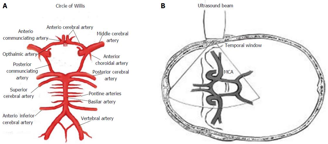
Figure 1 Circle of Willis and Ultrasonographic study by transcranial Doppler ultrasound.
A: Circle of Willis; B: Transmission of ultrasound beam through skull using pulsed Doppler sectorial probe with a 2.0-3.5 MHz emission frequency. Probe is positioned on temporal window. MCA: Middle cerebral artery.
- Citation: D’Andrea A, Conte M, Cavallaro M, Scarafile R, Riegler L, Cocchia R, Pezzullo E, Carbone A, Natale F, Santoro G, Caso P, Russo MG, Bossone E, Calabrò R. Transcranial Doppler ultrasonography: From methodology to major clinical applications. World J Cardiol 2016; 8(7): 383-400
- URL: https://www.wjgnet.com/1949-8462/full/v8/i7/383.htm
- DOI: https://dx.doi.org/10.4330/wjc.v8.i7.383


