Copyright
©The Author(s) 2016.
World J Cardiol. May 26, 2016; 8(5): 340-350
Published online May 26, 2016. doi: 10.4330/wjc.v8.i5.340
Published online May 26, 2016. doi: 10.4330/wjc.v8.i5.340
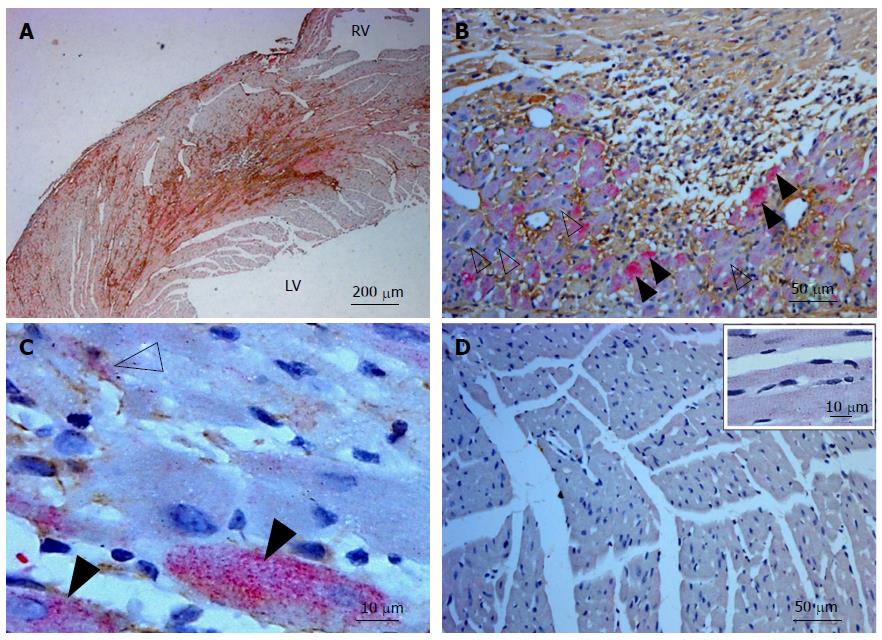
Figure 1 Tenascin C and toll-like receptor 4 staining in the murine left ventricle following infarction.
A: Low power view of the infarcted mouse LV showing the proximity of TNC (brown) and TLR4 (pink) 3 d following the occlusion of the left anterior coronary artery; B: Diffuse TLR4 staining (open arrows) in myocytes and interstitial cells and intense TLR4 staining (solid arrows) in some myocytes. Interstitial TNC staining (brown) is evident around these cells; C: High powered view of TLR4 (pink) and alpha smooth muscle actin (brown) staining of cells in the infarcted LV. Intense TLR4 staining can be seen in some myocytes (solid arrow) with more diffuse staining seen in some cardiac myofibroblasts (labelled both pink and brown, open arrow); D: Low power view of the non-infarcted side of the mouse myocardium stained for TNC (brown) and TLR4 (pink). An absence of TNC staining and light diffuse TLR4 staining of the cells is observed. Inset image: a high powered view of cells in this area. In each image cell nuclei were stained with Mayer’s Haematoxylin. RV: Right ventricle. LV: Left ventricle; TNC: Tenascin C; TLR4: Toll-like receptor 4.
- Citation: Maqbool A, Spary EJ, Manfield IW, Ruhmann M, Zuliani-Alvarez L, Gamboa-Esteves FO, Porter KE, Drinkhill MJ, Midwood KS, Turner NA. Tenascin C upregulates interleukin-6 expression in human cardiac myofibroblasts via toll-like receptor 4. World J Cardiol 2016; 8(5): 340-350
- URL: https://www.wjgnet.com/1949-8462/full/v8/i5/340.htm
- DOI: https://dx.doi.org/10.4330/wjc.v8.i5.340


