Copyright
©The Author(s) 2016.
World J Cardiol. Apr 26, 2016; 8(4): 310-316
Published online Apr 26, 2016. doi: 10.4330/wjc.v8.i4.310
Published online Apr 26, 2016. doi: 10.4330/wjc.v8.i4.310
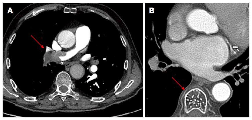
Figure 2 Examples of collateral findings detected with the preprocedural cardiac computed tomography.
A: Pulmonary thromboembolism involving principal branch of right pulmonary artery (red arrow); B: Classic “polka dotted” appearance due to the thickened vertebral trabeculae, highly suspicious for vertebral hemangioma (red arrow).
- Citation: Perna F, Casella M, Narducci ML, Dello Russo A, Bencardino G, Pontone G, Pelargonio G, Andreini D, Vitulano N, Pizzamiglio F, Conte E, Crea F, Tondo C. Collateral findings during computed tomography scan for atrial fibrillation ablation: Let’s take a look around. World J Cardiol 2016; 8(4): 310-316
- URL: https://www.wjgnet.com/1949-8462/full/v8/i4/310.htm
- DOI: https://dx.doi.org/10.4330/wjc.v8.i4.310


