Copyright
©The Author(s) 2016.
World J Cardiol. Apr 26, 2016; 8(4): 310-316
Published online Apr 26, 2016. doi: 10.4330/wjc.v8.i4.310
Published online Apr 26, 2016. doi: 10.4330/wjc.v8.i4.310
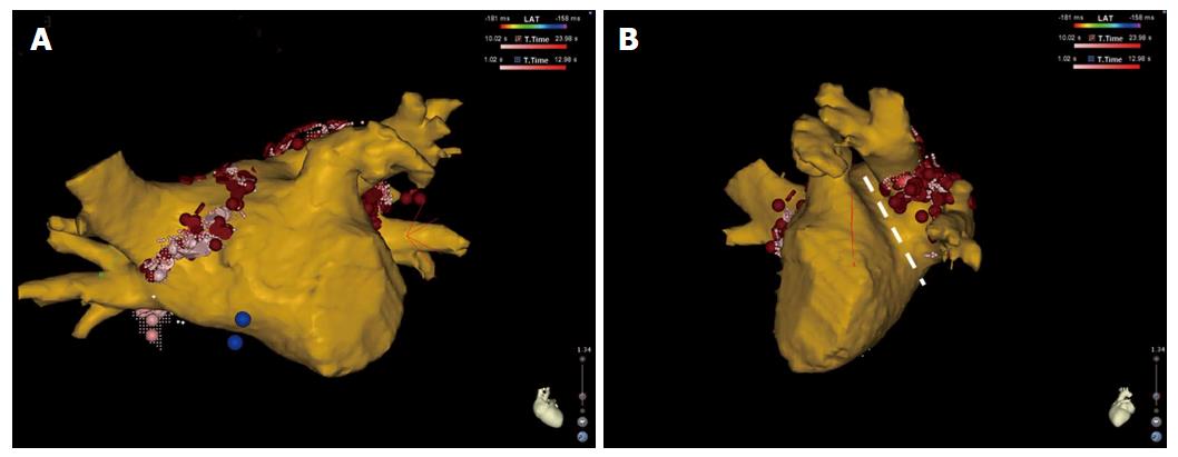
Figure 1 Antero-posterior (A) and left lateral (B) view of three-dimensional reconstruction of the left atrium and pulmonary veins after merging the cardiac computed tomography with the electroanatomical map created with the Carto 3 system (Biosense Webster, Inc.
, Diamond Bar, CA, United States). Note the ablation tags (red dots) placed around the pulmonary vein ostia on the computed tomography reconstruction. In the left lateral view, the narrow ridge between the left-sided pulmonary veins and the left atrial appendage can be appreciated (white dashed line).
- Citation: Perna F, Casella M, Narducci ML, Dello Russo A, Bencardino G, Pontone G, Pelargonio G, Andreini D, Vitulano N, Pizzamiglio F, Conte E, Crea F, Tondo C. Collateral findings during computed tomography scan for atrial fibrillation ablation: Let’s take a look around. World J Cardiol 2016; 8(4): 310-316
- URL: https://www.wjgnet.com/1949-8462/full/v8/i4/310.htm
- DOI: https://dx.doi.org/10.4330/wjc.v8.i4.310


