Copyright
©The Author(s) 2016.
World J Cardiol. Feb 26, 2016; 8(2): 146-162
Published online Feb 26, 2016. doi: 10.4330/wjc.v8.i2.146
Published online Feb 26, 2016. doi: 10.4330/wjc.v8.i2.146
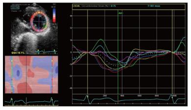
Figure 5 Two dimensional circumferential strain in a normal patient.
It represents left ventricle myocardial fiber shortening along the circular perimeter on a short-axis view and during systole, it is represented by synchronous negative curves.
- Citation: Rosa I, Marini C, Stella S, Ancona F, Spartera M, Margonato A, Agricola E. Mechanical dyssynchrony and deformation imaging in patients with functional mitral regurgitation. World J Cardiol 2016; 8(2): 146-162
- URL: https://www.wjgnet.com/1949-8462/full/v8/i2/146.htm
- DOI: https://dx.doi.org/10.4330/wjc.v8.i2.146


