Copyright
©The Author(s) 2016.
World J Cardiol. Feb 26, 2016; 8(2): 132-145
Published online Feb 26, 2016. doi: 10.4330/wjc.v8.i2.132
Published online Feb 26, 2016. doi: 10.4330/wjc.v8.i2.132
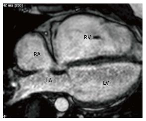
Figure 8 Arrhythmogenic right ventricular dysplasia.
Four-chamber cine steady state free precession image shows wall shows aneurysmal dilation of the right ventricle. There was also low ejection fraction (ejection fraction-35%) and severe right ventricle dilation (end diastolic volume index-130 mL/m2). These magnetic resonance imaging features satisfy one major criterion for arrhythmogenic right ventricular dysplasia. RA: Right atrium; LA: Left atrium; RV: Right ventricular; LV: Left ventricular.
- Citation: Kalisz K, Rajiah P. Impact of cardiac magnetic resonance imaging in non-ischemic cardiomyopathies. World J Cardiol 2016; 8(2): 132-145
- URL: https://www.wjgnet.com/1949-8462/full/v8/i2/132.htm
- DOI: https://dx.doi.org/10.4330/wjc.v8.i2.132


