Copyright
©The Author(s) 2016.
World J Cardiol. Feb 26, 2016; 8(2): 132-145
Published online Feb 26, 2016. doi: 10.4330/wjc.v8.i2.132
Published online Feb 26, 2016. doi: 10.4330/wjc.v8.i2.132
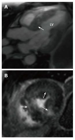
Figure 3 Hypertrophic cardiomyopathy.
A: Three-chamber steady state free precession image shows severe hypertrophy of the basal anteroseptum (arrow), which causes LVOT obstruction; B: Short-axis delayed enhancement image shows patchy mid myocardial enhancement in hypertrophied segments, suggestive of interstitial fibrosis in a pattern specific for hypertrophic cardiomyopathy. LV: Left ventricular.
- Citation: Kalisz K, Rajiah P. Impact of cardiac magnetic resonance imaging in non-ischemic cardiomyopathies. World J Cardiol 2016; 8(2): 132-145
- URL: https://www.wjgnet.com/1949-8462/full/v8/i2/132.htm
- DOI: https://dx.doi.org/10.4330/wjc.v8.i2.132


