Copyright
©The Author(s) 2016.
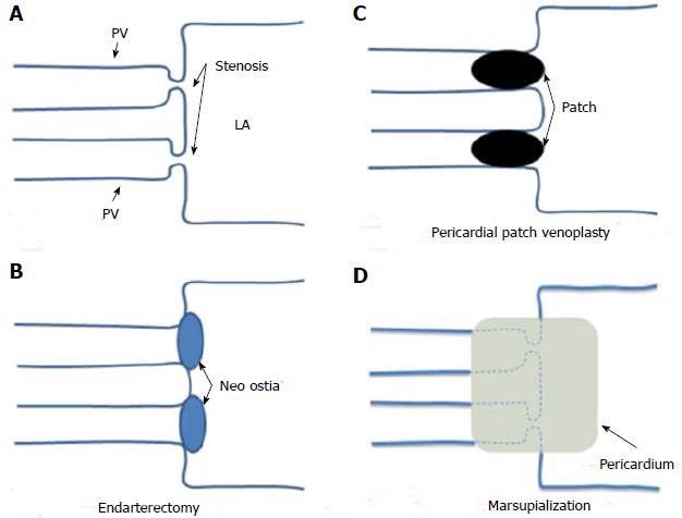
Figure 5 Surgical techniques for pulmonary veins.
A: Schematic representation of a bilateral pulmonary vein stenosis at the ostia of the vessels; B: Endarterectomy; the stenotic tissue has been excised and the PVs directly anastomosed to the LA; C: Pericardial patch venoplasty; the stenotic tissue has been resected and a pericardial patch anastomosis has been used to enlarge the tightened ostia of the vessels; D: Sutureless marsupialization: the veins ostia have been incised longitudinally, excess fibrotic tissue has been excised and in situ pericardial flaps have been sewn directly to the left atrium so direct stiches over the cut edges of the pulmonary veins are avoid. PV: Pulmonary vein; LA: Left atrium.
- Citation: Pazos-López P, García-Rodríguez C, Guitián-González A, Paredes-Galán E, Álvarez-Moure M&DLG, Rodríguez-Álvarez M, Baz-Alonso JA, Teijeira-Fernández E, Calvo-Iglesias FE, Íñiguez-Romo A. Pulmonary vein stenosis: Etiology, diagnosis and management. World J Cardiol 2016; 8(1): 81-88
- URL: https://www.wjgnet.com/1949-8462/full/v8/i1/81.htm
- DOI: https://dx.doi.org/10.4330/wjc.v8.i1.81


