Copyright
©The Author(s) 2016.
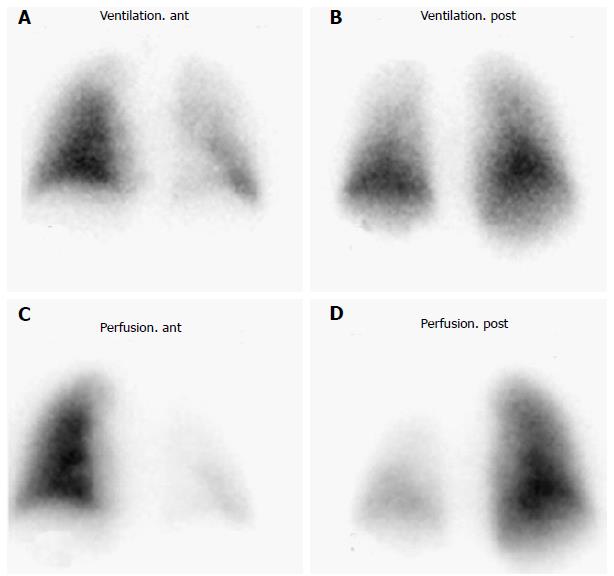
Figure 4 Radionuclide lung ventilation/perfusion scan performed three months after radiofrequency ablation in a patient with shortness of breath.
A and B: Normal ventilation; C and D marked hypoperfusion within the left lung consistent with significant left PV stenosis which was demonstrated on a CT scan. PV: Pulmonary vein; CT: Computed tomography.
- Citation: Pazos-López P, García-Rodríguez C, Guitián-González A, Paredes-Galán E, Álvarez-Moure M&DLG, Rodríguez-Álvarez M, Baz-Alonso JA, Teijeira-Fernández E, Calvo-Iglesias FE, Íñiguez-Romo A. Pulmonary vein stenosis: Etiology, diagnosis and management. World J Cardiol 2016; 8(1): 81-88
- URL: https://www.wjgnet.com/1949-8462/full/v8/i1/81.htm
- DOI: https://dx.doi.org/10.4330/wjc.v8.i1.81


