Copyright
©The Author(s) 2016.
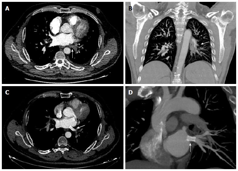
Figure 2 Computed tomography of a patient undergone radiofrequency ablation two months before and recent onset of dyspnea on exertion.
A: Absent of contrast (arrow) in the left lower pulmonary vein (complete occlusion); B: Extensive infiltrate within the left lung (arrow) cause by localized edema; C and D: After stent implantation (arrows) flow was successfully restored.
- Citation: Pazos-López P, García-Rodríguez C, Guitián-González A, Paredes-Galán E, Álvarez-Moure M&DLG, Rodríguez-Álvarez M, Baz-Alonso JA, Teijeira-Fernández E, Calvo-Iglesias FE, Íñiguez-Romo A. Pulmonary vein stenosis: Etiology, diagnosis and management. World J Cardiol 2016; 8(1): 81-88
- URL: https://www.wjgnet.com/1949-8462/full/v8/i1/81.htm
- DOI: https://dx.doi.org/10.4330/wjc.v8.i1.81


