Copyright
©The Author(s) 2015.
World J Cardiol. Sep 26, 2015; 7(9): 579-582
Published online Sep 26, 2015. doi: 10.4330/wjc.v7.i9.579
Published online Sep 26, 2015. doi: 10.4330/wjc.v7.i9.579
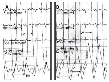
Figure 4 Hemodynamic tracing during catheterization.
A: Right atrial (RA) pressure tracing shows prominent X and Y descent; B: Ventricular pressure tracing shows typical “dip-and-plateau configuration” during diastole. RV: Right ventricle; LV: Left ventricle.
- Citation: Vijayvergiya R, Vadivelu R, Mahajan S, Rana SS, Singhal M. Eggshell calcification of the heart in constrictive pericarditis. World J Cardiol 2015; 7(9): 579-582
- URL: https://www.wjgnet.com/1949-8462/full/v7/i9/579.htm
- DOI: https://dx.doi.org/10.4330/wjc.v7.i9.579


