Copyright
©The Author(s) 2015.
World J Cardiol. Sep 26, 2015; 7(9): 579-582
Published online Sep 26, 2015. doi: 10.4330/wjc.v7.i9.579
Published online Sep 26, 2015. doi: 10.4330/wjc.v7.i9.579
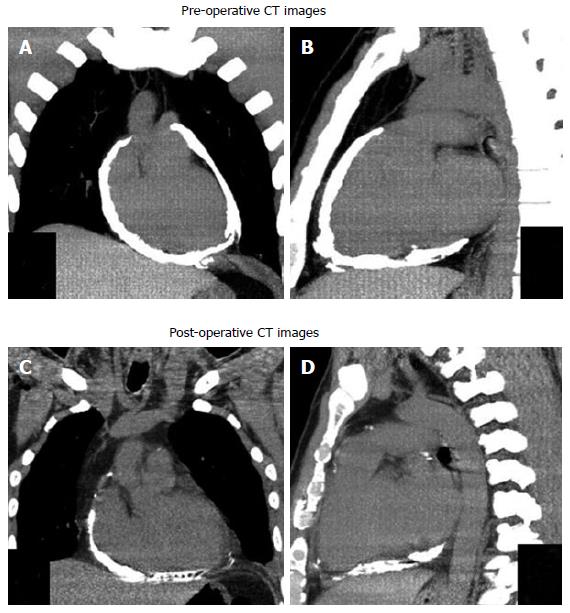
Figure 2 Computed tomography.
Non-contrast computed tomography reconstructed images in coronal (A) and sagittal (B) planes show thick, calcified pericardium around the heart. A repeat CT following partial pericardiectomy in coronal (C) and sagittal (D) planes show residual calcified pericardium at right atrial and posterior surface of the heart. Calcified pericardium is absent along the antero-lateral surface of the heart. CT: Computed tomography.
- Citation: Vijayvergiya R, Vadivelu R, Mahajan S, Rana SS, Singhal M. Eggshell calcification of the heart in constrictive pericarditis. World J Cardiol 2015; 7(9): 579-582
- URL: https://www.wjgnet.com/1949-8462/full/v7/i9/579.htm
- DOI: https://dx.doi.org/10.4330/wjc.v7.i9.579


