Copyright
©The Author(s) 2015.
World J Cardiol. Aug 26, 2015; 7(8): 499-503
Published online Aug 26, 2015. doi: 10.4330/wjc.v7.i8.499
Published online Aug 26, 2015. doi: 10.4330/wjc.v7.i8.499
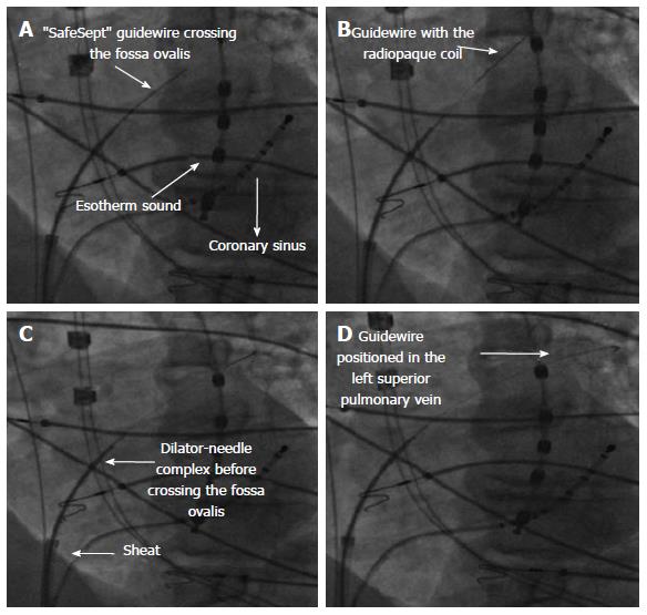
Figure 3 Sequence of fluoroscopy imaging of trans-septal puncture in the same projection (left anterior oblique view).
A: The esotherm sound for the esophageal temperature control and coronary sinus catheter are in place. The “SafeSept” guidewire penetrates the fossa ovalis; B: The “SafeSept” guidewire is visible in the left atrium thanks to radiopaque coil; C: The trans-septal assembly (needle, dilator and sheath) is placed in the right atrium before crossing the fossa ovalis; D: The distal part of trans-septal guidewire is positioned in the left superior pulmonary vein.
- Citation: Zucchetti M, Casella M, Russo AD, Fassini G, Carbucicchio C, Russo E, Marino V, Catto V, Tondo C. Difficult case of a trans-septal puncture: Use of a “SafeSept” guidewire. World J Cardiol 2015; 7(8): 499-503
- URL: https://www.wjgnet.com/1949-8462/full/v7/i8/499.htm
- DOI: https://dx.doi.org/10.4330/wjc.v7.i8.499


