Copyright
©The Author(s) 2015.
World J Cardiol. Jul 26, 2015; 7(7): 431-433
Published online Jul 26, 2015. doi: 10.4330/wjc.v7.i7.431
Published online Jul 26, 2015. doi: 10.4330/wjc.v7.i7.431
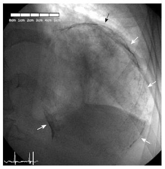
Figure 5 Fluoroscopic imaging in right anterior oblique projection showing a complete oval calcified mass (arrows), corresponding with the left ventricular aneurysm.
- Citation: de Agustin JA, de Diego JJG, Marcos-Alberca P, Rodrigo JL, Almeria C, Mahia P, Luaces M, Garcia-Fernandez MA, Macaya C, de Isla LP. Giant and thrombosed left ventricular aneurysm. World J Cardiol 2015; 7(7): 431-433
- URL: https://www.wjgnet.com/1949-8462/full/v7/i7/431.htm
- DOI: https://dx.doi.org/10.4330/wjc.v7.i7.431


