Copyright
©The Author(s) 2015.
World J Cardiol. Jun 26, 2015; 7(6): 361-366
Published online Jun 26, 2015. doi: 10.4330/wjc.v7.i6.361
Published online Jun 26, 2015. doi: 10.4330/wjc.v7.i6.361
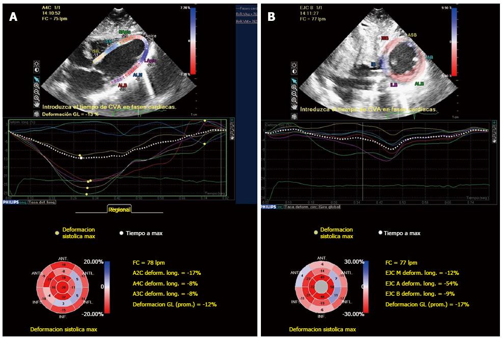
Figure 4 Bull’s eye mapping of two-dimensional speckle tracking strain imaging longitudinal (A) and circunferencial (B) showed decreased strain values of the basal and mid-ventricular segments, with normal or increased strain values of the apical segments.
- Citation: Robles P, Monedero I, Rubio A, Botas J. Reverse or inverted apical ballooning in a case of refeeding syndrome. World J Cardiol 2015; 7(6): 361-366
- URL: https://www.wjgnet.com/1949-8462/full/v7/i6/361.htm
- DOI: https://dx.doi.org/10.4330/wjc.v7.i6.361


