Copyright
©The Author(s) 2015.
World J Cardiol. Jun 26, 2015; 7(6): 361-366
Published online Jun 26, 2015. doi: 10.4330/wjc.v7.i6.361
Published online Jun 26, 2015. doi: 10.4330/wjc.v7.i6.361
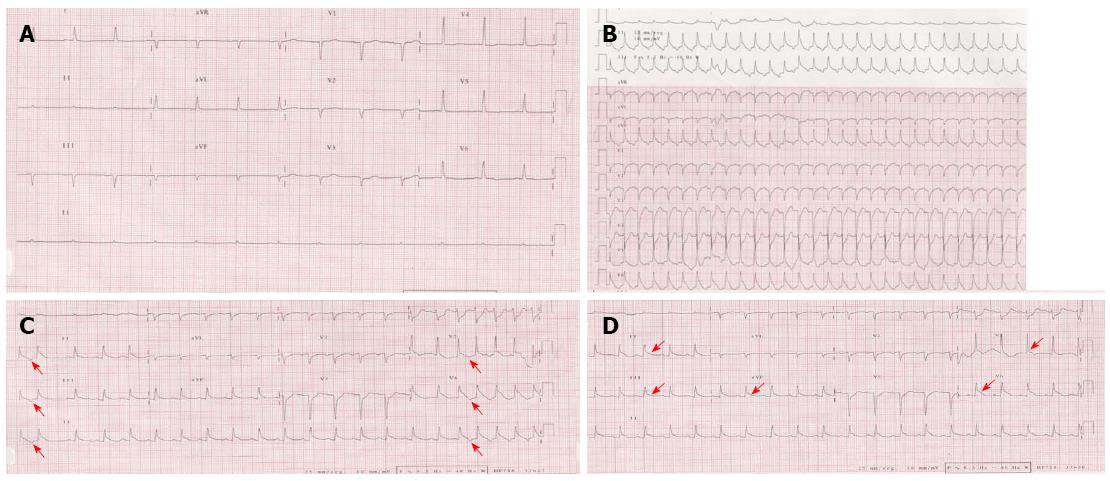
Figure 1 Change process of electrocardiogram.
Baseline electrocardiogram (ECG) showed sinus bradycardia and nonspecific repolarization abnormalities (A). Surface ECG of repetitive nonsustained atrial tachycardia (AT). Note that the first P wave of the tachycardia is similar in morphology to the subsequent P waves, consistent with abnormal automaticity as the mechanism of the AT (red arrow). In the setting of posteroseptal AT (originating below and around the coronary sinus ostium), the P wave is positive in lead V1, negative in the inferior leads, and positive in leads aVL and aVR (B and C). ECG after the completion of the tachycardia showed persistent ST elevation in leads II, III, AVF, V5 and V6 (D) (red arrows).
- Citation: Robles P, Monedero I, Rubio A, Botas J. Reverse or inverted apical ballooning in a case of refeeding syndrome. World J Cardiol 2015; 7(6): 361-366
- URL: https://www.wjgnet.com/1949-8462/full/v7/i6/361.htm
- DOI: https://dx.doi.org/10.4330/wjc.v7.i6.361


