Copyright
©The Author(s) 2015.
World J Cardiol. Apr 26, 2015; 7(4): 224-229
Published online Apr 26, 2015. doi: 10.4330/wjc.v7.i4.224
Published online Apr 26, 2015. doi: 10.4330/wjc.v7.i4.224
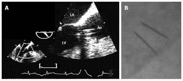
Figure 1 Transesophageal echo and fluoroscopy.
A: Transesophageal Echo: 3-chamber view at 129°, with zoom in the LV outflow tract, showing the prosthetic valve with its parallel discs; B: Fluoroscopy showing the almost parallel tilting discs. Both exams confirm an adequate prosthetic valve opening. LA: Left atrium; LV: Left ventricle; Ao: Aorta.
- Citation: Soumoulou JB, Cianciulli TF, Zappi A, Cozzarin A, Saccheri MC, Lax JA, Guidoin R, Zhang Z. Limitations of multimodality imaging in the diagnosis of pannus formation in prosthetic aortic valve and review of the literature. World J Cardiol 2015; 7(4): 224-229
- URL: https://www.wjgnet.com/1949-8462/full/v7/i4/224.htm
- DOI: https://dx.doi.org/10.4330/wjc.v7.i4.224


