Copyright
©The Author(s) 2015.
World J Cardiol. Apr 26, 2015; 7(4): 167-177
Published online Apr 26, 2015. doi: 10.4330/wjc.v7.i4.167
Published online Apr 26, 2015. doi: 10.4330/wjc.v7.i4.167
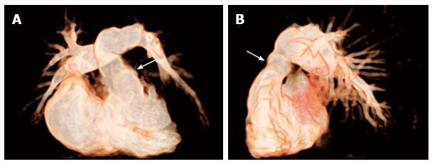
Figure 4 Non-invasive 3D whole heart imaging by magnetic resonance tomography.
Non-invasive 3D whole heart imaging by magnetic resonance tomography was performed in a patient with pulmonary atresia with intact ventricular septum after repair by pulmonic homograft implantation (arrows) with RVOT dysfunction prior to PPVI (A) in a.p. and (B) in lat. view. (Courtesy of Wagner R). RVOT: Right ventricular outflow tract; PPVI: Percutaneous pulmonary valve implantation.
- Citation: Wagner R, Daehnert I, Lurz P. Percutaneous pulmonary and tricuspid valve implantations: An update. World J Cardiol 2015; 7(4): 167-177
- URL: https://www.wjgnet.com/1949-8462/full/v7/i4/167.htm
- DOI: https://dx.doi.org/10.4330/wjc.v7.i4.167


