Copyright
©The Author(s) 2015.
World J Cardiol. Mar 26, 2015; 7(3): 157-160
Published online Mar 26, 2015. doi: 10.4330/wjc.v7.i3.157
Published online Mar 26, 2015. doi: 10.4330/wjc.v7.i3.157
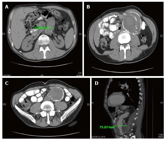
Figure 1 Computed tomography scan.
A: Showing aortic abdominal aneurysm at the level of renal arteries; B: Showing massive unruptured abdominal aortic aneurysm along with loops of small intestine; C: Showing continuation of abdominal aortic aneurysm along with plaques of calcification surrounding it; D: Showing longitudinal section of contrast filled abdominal aortic aneurysm with bilateral mural thrombus.
- Citation: Saade C, Pandya B, Raza M, Meghani M, Asti D, Ghavami F. 9.1 cm abdominal aortic aneurysm in a 69-year-old male patient. World J Cardiol 2015; 7(3): 157-160
- URL: https://www.wjgnet.com/1949-8462/full/v7/i3/157.htm
- DOI: https://dx.doi.org/10.4330/wjc.v7.i3.157


