Copyright
©The Author(s) 2015.
World J Cardiol. Feb 26, 2015; 7(2): 86-100
Published online Feb 26, 2015. doi: 10.4330/wjc.v7.i2.86
Published online Feb 26, 2015. doi: 10.4330/wjc.v7.i2.86
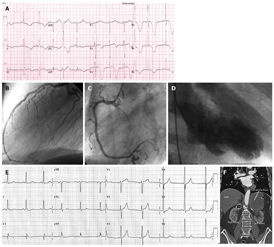
Figure 7 Patient 7.
A: An electrocardiographic (ECG) tracing, showing giant T wave inversion in the precordial leads V2-6, of a 76-year Caucasian male with a past medical history of an old inferior myocardial infarction, percutaneous coronary intervention of the right coronary artery (RCA) and left anterior descending coronary artery, acutely presented with left abdominal pain, psychomotor unrest, diaphoresis and blood pressure difference between the right and left arm. Acute aortic dissection was excluded as well as recurrent MI. Coronary angiography frame of (B) the left coronary artery and (C) the RCA depicting no significant stenosis of the arterial tree. Serum cardiac markers were slightly elevated. Echocardiographic (hypokinesia of the mid and apical regions and hyperkinesia of the basal segments) findings and (D) ventriculography (apical ballooning) were all compatible with Takotsubo cardiomyopathy; E: Base line ECG. The abdominal ultrasound and (F) CT demonstrated a pheochromocytoma in the left adrenal region which was confirmed with 123I-MIBG scan and dihydroxyphenylalanine-Positron Emission tomography and proved by pathological results. Plasma and urine metanephrin and normetanephrin were highly elevated. After removal of the hormonally active tumor, the patient became symptom free and the ECG normalized. The medical treatment continued including calcium reentry blocker, beta blocker, aspirin, angiotensin converting enzyme inhibitor, statin and an α-blocker.
- Citation: Said SA, Bloo R, Nooijer R, Slootweg A. Cardiac and non-cardiac causes of T-wave inversion in the precordial leads in adult subjects: A Dutch case series and review of the literature. World J Cardiol 2015; 7(2): 86-100
- URL: https://www.wjgnet.com/1949-8462/full/v7/i2/86.htm
- DOI: https://dx.doi.org/10.4330/wjc.v7.i2.86


