Copyright
©The Author(s) 2015.
World J Cardiol. Feb 26, 2015; 7(2): 86-100
Published online Feb 26, 2015. doi: 10.4330/wjc.v7.i2.86
Published online Feb 26, 2015. doi: 10.4330/wjc.v7.i2.86
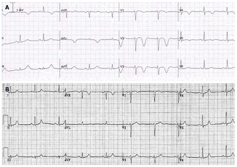
Figure 4 Patient 4.
A: An electrocardiographic (ECG) tracing, demonstrating negative T wave in the precordial leads V2-5, of a 69-year female patient with past medical history of transient ischemic attack two years previously, presented with interscapular pain. She had no emotional or physical stress. Normal results were found on transthoracic echocardiography, perfusion-ventilation scintigraphy, Coronary angiography and cardiac MRI. Brain CT scan revealed mild cerebral atrophy and minimal ischemic changes; B: The ECG showed spontaneous regression in 2 mo time. The etiology of the negative T wave inversion remains undetermined. Her medical regimen included aspirin, beta blocker, statin and diuretic.
- Citation: Said SA, Bloo R, Nooijer R, Slootweg A. Cardiac and non-cardiac causes of T-wave inversion in the precordial leads in adult subjects: A Dutch case series and review of the literature. World J Cardiol 2015; 7(2): 86-100
- URL: https://www.wjgnet.com/1949-8462/full/v7/i2/86.htm
- DOI: https://dx.doi.org/10.4330/wjc.v7.i2.86


